
You have goat to be kidding meA 1-month old Boer buckling was presented to the veterinarian for lethargy, recumbence, distended abdomen, tachycardia, and hypoglycemia. He was diagnosed with metabolic acidosis and was unable to recover. During necropsy several of the following specimens were recovered from hair. Video 1: Specimens recovered from the hair. Linognathus africanus (African goat louse) are Anoplurans or bloodsucking lice, with piercing mouthparts used for feeding. They spend their time on the hairs of the host, where they deposit eggs. All life stages occur on the host. Linognathus africanus and L. stenopsis, are species that can infest goats. Linognathus spp. first pair of legs is shorter than the second and third pairs, and L. africanus have a greatly expanded head behind the antennae compared to L. stenopsis. The African goat louse is distributed world-wide. Infestations by these lice are usually localized around the head, while the goat sucking louse (L. stenopsis) can be dispersed over the entire body. Lice are usually a nuisance, but in some cases severe infestations can cause scabby, and bleeding areas that may lead to bacterial infections. Pawsitive fecalA fecal sample from a 2-year old domestic shorthair cat was submitted to the parasitology laboratory for a wellness checkup. The fecal sample was normal in color and consistency. Fecal centrifugation with Sheather’s sugar solution revealed the presence of the following eggs: 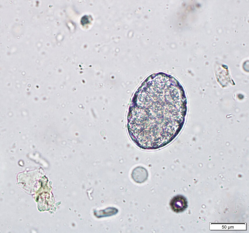 Image 1: Parasite identified on fecal flotation measuring 57 x 40 µM. Ancylostoma tubaeforme or cat hookworm. Infection with this parasite occurs when cats ingest infected eggs from the environment, through skin penetration by infected larvae, or by ingestion of a paratenic host. Unlike dogs, transmammary transmission does not occur and infection in cats is not as pathogenic as in dogs either. Adults live in the small intestine where they ingest blood from their host. Clinical signs include weight loss and regenerative anemia. The case of the unlucky lizardA bearded dragon was presented to a Veterinary Teaching Hospital due to lethargy and crusting of the eyes. A fecal analysis was requested as part of the clinical examination. The fecal flotation with Sheather’s sugar solution revealed the following structure:  Image 1: Parasite identified on fecal flotation measuring 22 x 24 µM. Isospora amphiboluri
A common coccidian found in captive bearded dragons (Pogona vitticeps) commercially bred. It infects enterocytes of the small and large intestine. In experimental infections, gross changes in the feces have not been observed, however young lizards can be severely affected with up to 15% mortality. The prepatent period is between 2 to 3 weeks, which is essential to consider when introducing new animals into a colony. The oocysts are 22.7 to 25.4 µM in diameter with a bilayer wall. The micropyle is absent and the sporulated oocysts contain two sporocysts measuring 11 to 17 µM. The sporocysts are single layered with a smooth wall. Roses are red, violets are blue, what's lurking inside you?A fecal sample from a 6-month old golden hamster, initially presented for rectal prolapse and surgery, was submitted to the parasitology diagnostics laboratory to evaluate the presence of parasites eggs. The fecal sample was centrifuged with Sheather’s sugar solution and the following egg was observed. 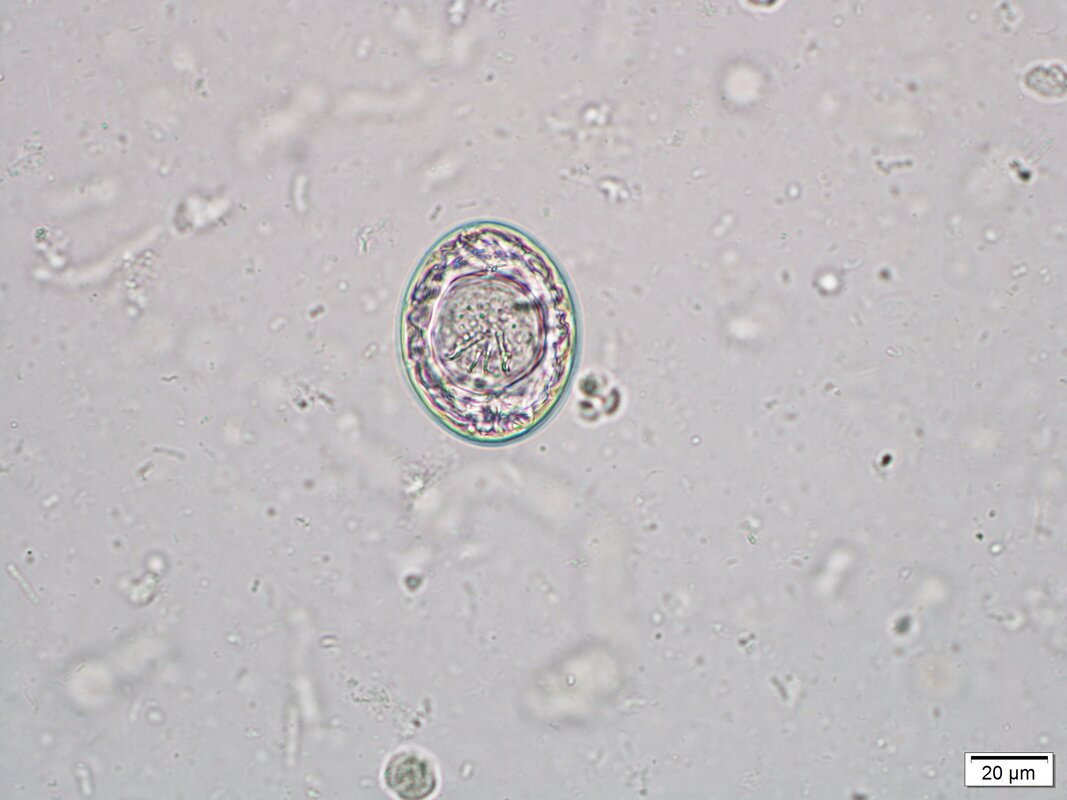 Image 1: Parasite egg measures 45 µm x 30 µm. Diagnosis: Hymenolepis nana, a parasite of rodents and humans. Egg size ranges from 30 to 50 µm. The inner membrane has two poles, from which 4-8 polar filaments spread out between the two membranes. The oncosphere has six hooks. The infective larval stage called Cysticercoid, develops in fleas and flour beetles. Besides these intermediate hosts, H. nana can complete its life cycle within the intestinal tract of a rodent or human, where the eggs hatch and form a cysticercoid in the intestinal mucosa. These cysticercoids later reenter the lumen to complete their development. The importance of this parasite is the zoonotic potential. New Year, New ProblemA litter of three Labrador retriever mix puppies were rescued from a local shelter in Oklahoma. As part of the clinical exam a fecal analysis was performed. The following eggs and parasites were observed on fecal centrifugation with sugar Sheather’s solution (Images 1-3). 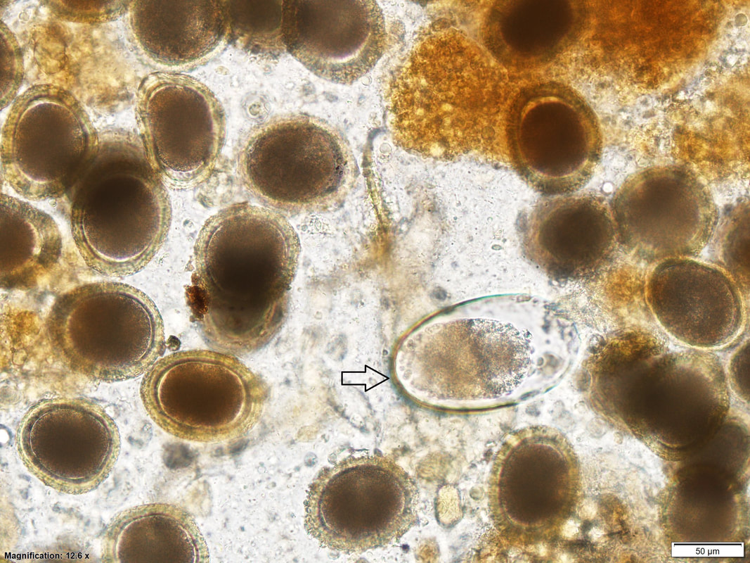 Image 1: Parasite eggs at 100x magnification. Arrow pointing to egg measuring 150 µM x 75 µM. 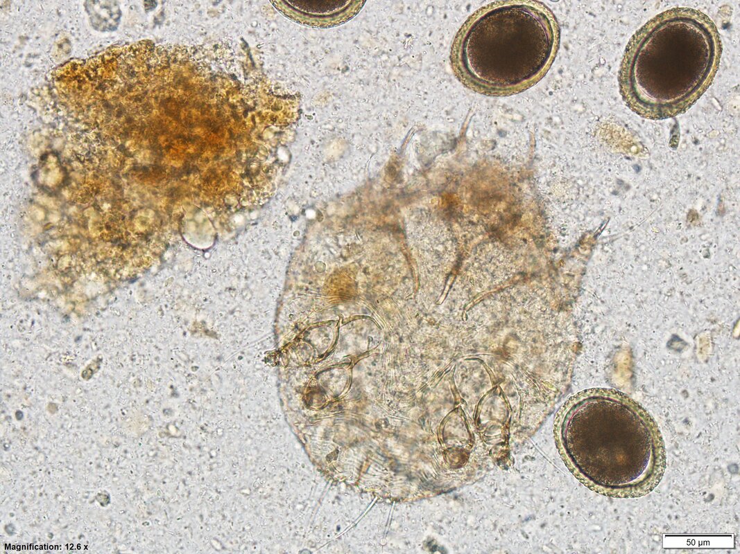 Image 2: 100x magnification. 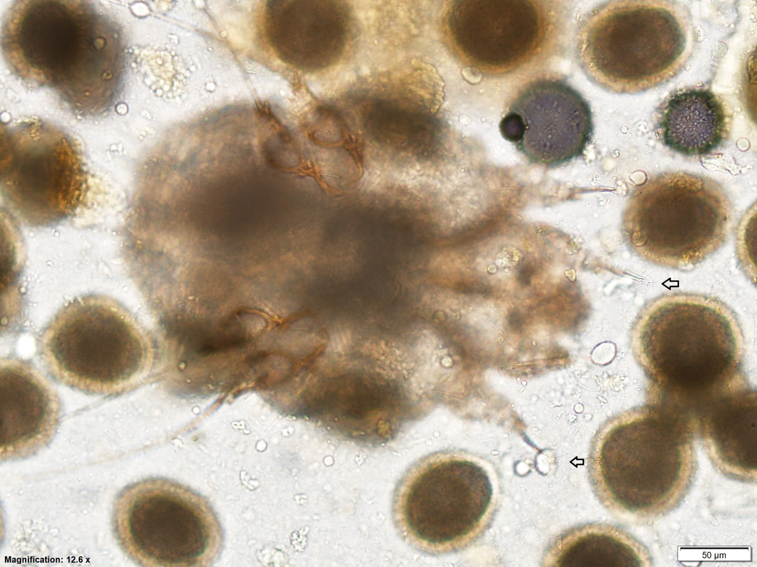 Image 3: Arrows pointing to distinct feature of the parasite. Sarcoptes scabiei, also called scabies mite in dogs. Sarcoptes are found in domestic animals and humans. They are transmitted by direct contact with an infested animal or contaminated fomites. Female mites burrow into the epidermis and deposit eggs. (Numerous Toxocara canis also observed on image.) Sarcoptes cause pruritus, alopecia and thickening of the skin. Although initially found on areas with thick skin or hairless, they can affect large areas of the skin. Skin scrapings are the diagnostic method, however mites can be difficult to recover. In some cases, mites are passed in the feces of the host after they ingest them while trying to relieve the intense pruritus. Female mites measure an average of 400 µM while males are smaller around 250 µM. They both have suckers on the unsegmented stalks on first and second pairs (image 4, small arrows) and males also have suckers on the fourth pair. The third and fourth pair of legs are short and rarely project past the margin of the body. All I want for Christmas is to be parasite free!A fecal sample from a 2 year-old cat with history of anorexia, was submitted to the Oklahoma Diagnostic Laboratory for fecal analysis. A fecal flotation with Zinc sulfate solution (specific gravity 1.18) was performed and the following eggs were observed (Photos 1-2). 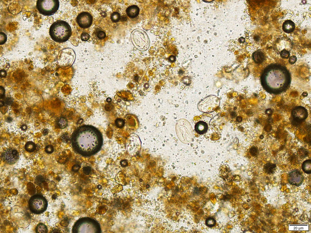 Photo 1: Parasite eggs at 100x magnification. Sizes range from 35 µM to 40 µM. 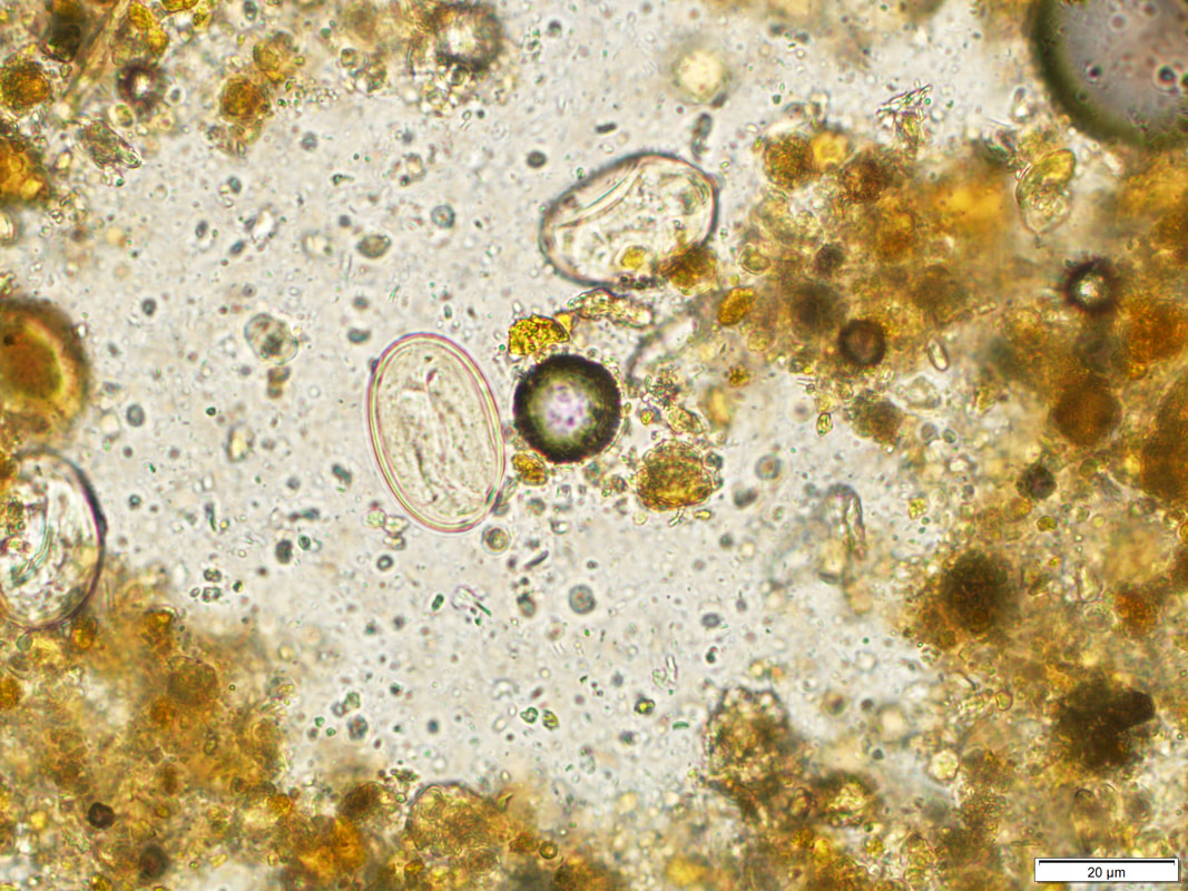 Photo 2: Closer view with larva inside the egg. Physalpotera spp.
Physalpotera spp. are Spirurid parasites of the stomach of dogs, cats and other wild mammals. Clinical signs are rare and may include vomiting and anorexia. The recommended detection method is fecal sedimentation (photo 3, below), however on occasion they can be detected with fecal centrifugation. Life cycle: The eggs are ingested by beetles, cockroaches or crickets where they develop into the infective stage. The definitive host in this case a cat, becomes infected when ingests the intermediate host or paratenic hosts such as reptiles. 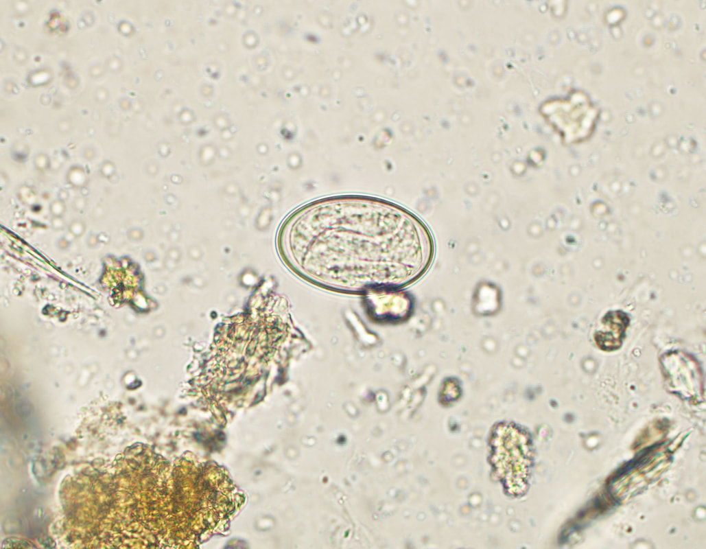
Photo 3: Physaloptera spp. recovered by sedimentation. |