
Dog"ged" microfilariaeA blood sample from a 21-month old female Boston Terrier Mix, was submitted to the parasitology diagnostic lab to be tested for Dirofilaria immitis detection. The dog had a history of microfilaremia in 2020 but no antigen was detected during that same year. In September 2021, during the annual wellness a snap test was performed at the clinic with no antigen detected, but microfilaria was again observed on the direct smear. To rule out the presence of antigen-antibody complexes, the serum sample was heat treated at the diagnostic lab, but no antigen was detected. The Modified Knott’s test showed the presence of the following microfilariae. (Images 1-3) Size average 275 µm in length x 5 µm in width. Images 1-3: Key features for identification. Acanthocheilonema reconditum, is a member of the subfamily Onchocercinae that infects canids. It is transmitted by fleas and the louse Heterodoxus spiniger. The adults are located in the subcutaneous tissues where they do not cause any pathology. The importance of this parasite lies on the confusion that its microfilariae can create on the diagnosis of Dirofilaria immitis infection. Both microfilariae are similar and can be easily confused, however morphological characteristics and motility can help differentiate these two microfilariae. Observing microfilariae in puppies and young dogs with no D. immitis antigen detection can be explained by infection of A. reconditum such as this case or transplacental transmission of D. immitis. Not another coughing catA 6-year-old formerly-feral domestic shorthair cat presented to a veterinary internist in Virginia for chronic coughing. Serum antigen and antibody testing for heartworm infection was negative. A fecal sample was collected and submitted to a diagnostic laboratory for evaluation, which revealed the presence of the following parasite ova (Image 1). In addition, thoracic radiographs were taken (Image 2). Based on these results the cat was treated by the internist with weekly fenbendazole and ivermectin, but the parasite persisted in serial fecal exams and recheck radiographs were relatively unchanged. 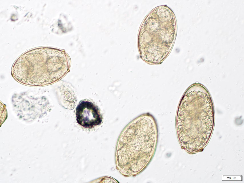 Image 1: Ova found on fecal examination (40x objective). 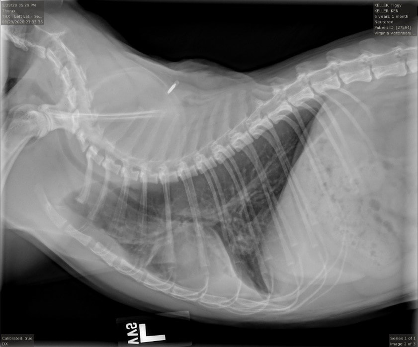 Image 2: Left lateral radiographic view of thorax. Radiographs demonstrated right middle lung lobe consolidation with heavy bronchial pattern and cavitated lesions. Paragonimus kellicotti, also known as the lung fluke, is a trematode found throughout North America. The parasite has a 3-host life cycle in which the infective larval stage (miricidium) first penetrates an aquatic snail, where it develops into a cercaria and later exits the snail to encyst within the tissues of crawfish. The life cycle is completed when a vertebrate definitive host (animal or human) consumes a crawfish containing these cysts, and the parasites migrate from the gastrointestinal tract to the lungs where they encyst and reproduce. Eggs are coughed up in sputum, swallowed, and passed into the environment via the host's feces. Encystment of Paragonimus within the lung parenchyma most often results in the clinical signs of chronic, persistent coughing (in rare cases, rupture of a lung cyst can result in pneumothorax and acute dyspnea). Large cysts may be appreciated on thoracic radiographs as nodular or cavitary lesions, particularly in the right caudal lung lobes. In this case, the paragonimiasis in this cat likely persisted due to an inadequate treatment protocol. Paragonimus treatment differs from that of lung nematodes in that it requires the use of praziquantel (often used in conjunction with a benzimidazole) in order to eliminate the parasite effectively. "Acarologic" ventureIn February 2021, this tick was removed from a 4-year-old male castrated Jack Russell Terrier from Texas, and sent to the parasitology lab for identification. The lab worker was able to take pictures showcasing key features for identifying this specimen, as seen below. 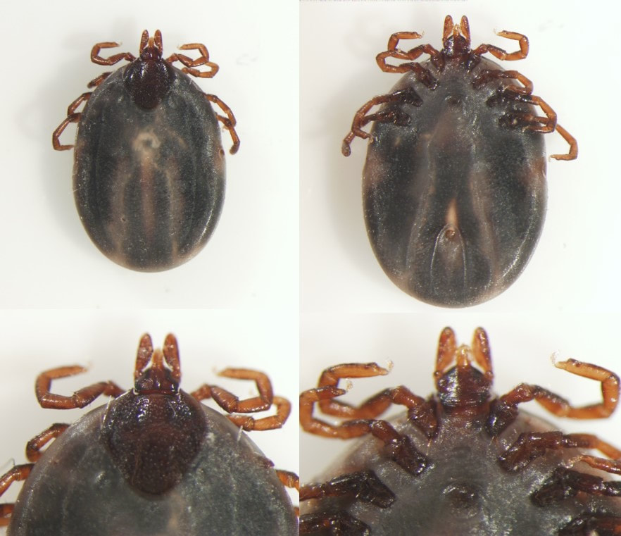 Image 1: Key features for identification. This is Ixodes cookei, the groundhog tick or woodchuck tick. Female Ixodes cookei can be differentiated from other Ixodes species based on their angular scutum shape and relatively short palps. Females of Ixodes scapularis will instead have a rounded, oval shaped scutum, and longer palps, as seen in Image 2 (right).
Groundhog ticks are found throughout eastern North America. In northeastern states, the upper Midwest, and eastern Canada, this tick serves as a vector for Powassan virus and may also transmit Borrelia burgdorferi. In Canada, adult Ixodes cookei are most active in the summer but in the southern US this tick is more commonly found in the winter months. 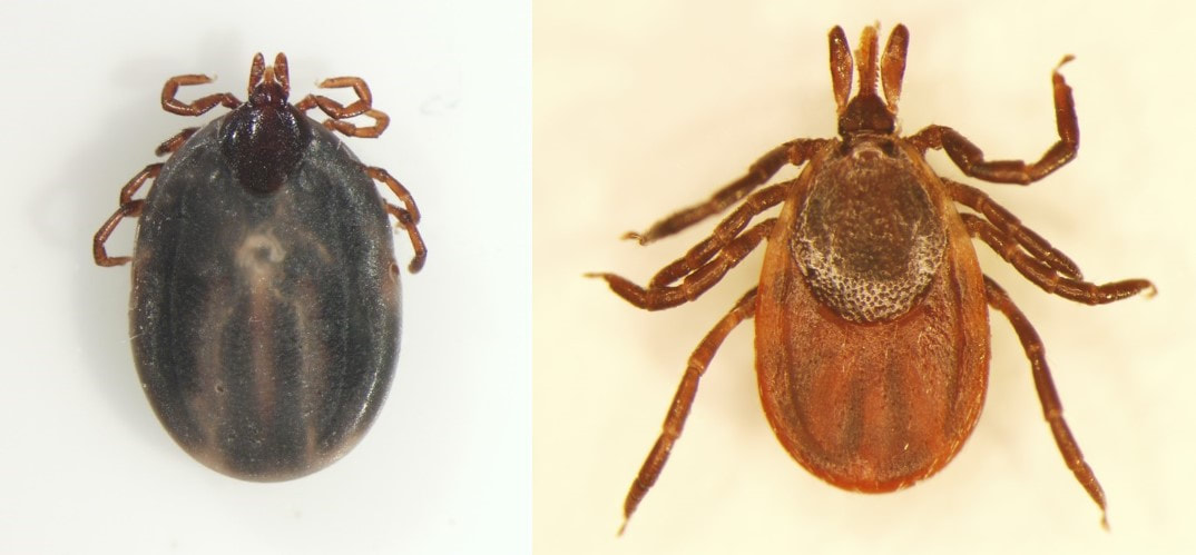
Image 2: Adult female Ixodes cookei (left) and Ixodes scapularis (right). Hooked on youAn adult female 3-year-old greyhound presented to a veterinarian in Illinois for persistent hookworm infection. She had been dewormed monthly with Pyrantel, Febantel, and transdermal Moxidectin for the past 3 months. A fecal sample was collected and a quantitative fecal egg count was performed using a modified McMaster test. The following eggs were found and counted at 850 eggs per gram (Image 1). 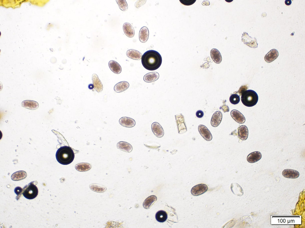 Image 1: Eggs found on fecal examination (10x objective). Multi-drug resistant Ancylostoma caninum is a growing concern and commonly reported in retired racing greyhounds although the issue has also been identified in other breeds of dogs. To confirm multi-drug resistance, a fecal egg count reduction test (FECRT) is performed by collecting samples immediately prior to treatment and again 14 days following treatment. If hookworms are susceptible to the anthelmintic used, a FECRT should show a reduction of >95%. Because this dog has already been dewormed with all three anthelmintic classes, resistance is likely and off-label use of Emodepside can be considered following all appropriate safety and regulatory recommendations (see reference). A FECRT should be performed to confirm efficacy. In addition, centrifugal fecal floats – which are more sensitive than modified McMaster test – should be performed monthly for 3 months to confirm the dog does not become reinfected. During this time, it is important to maintain strict fecal clean-up and avoid dog parks to minimize sources of reinfection until no eggs are consistently seen on fecal analysis. There are 4 routes of transmission for hookworms in dogs: ingestion of infective L3 larvae, direct penetration of larvae percutaneously, transmammary transmission from mother to pups, and ingestion of paratenic hosts containing the infective L3. An event that can be confused with persistent hookworm infection is "larval leak", when encysted larvae migrate from tissues and colonize the small intestine and becoming patent. Due to this migration, fecal egg counts 14 days after treatment are important. Earlier fecal egg counts may reflect egg suppression following treatment but not true efficacy, and if eggs are present in the feces 21 days or more after treatment, reinfection from the environment or via larval leak is suggested rather than resistance. What's on the hook?Several crappie fish were submitted from the Atoka Lake (Oklahoma) for necropsy due to lacerations observed by anglers. Samples were collected for parasitology testing, including fecal samples and the stomach of the fish. Examination of the stomach content revealed the following parasites in one of the fish (Image 1), and fecal centrifugation with Sheather’s sugar solution revealed the presence of larvated eggs (Image 2). 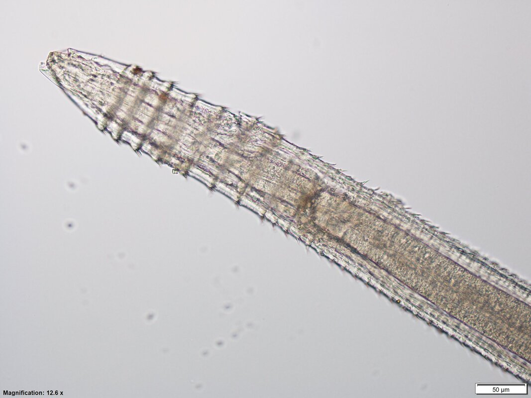 Image 1: Anterior end of parasite found in stomach (~7mm in length). 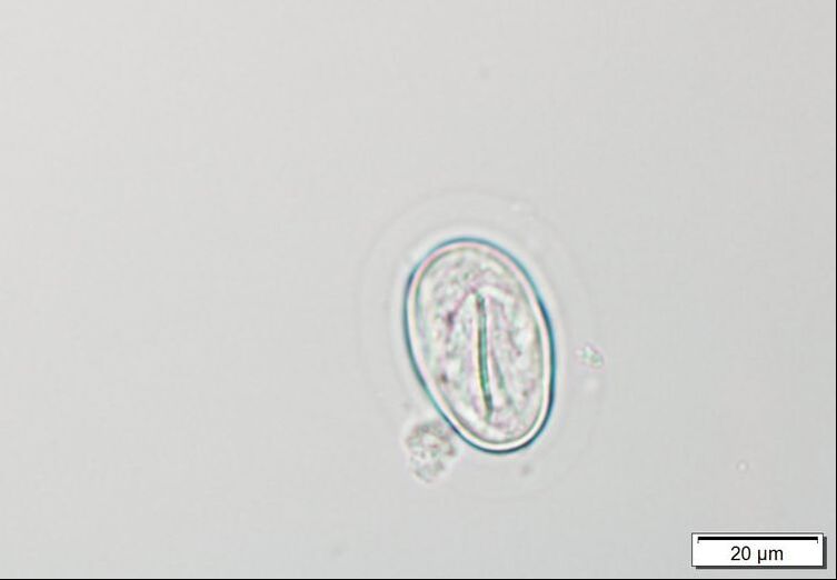 Image 2: Egg found in fecal flotation measuring 37.5 x 25µM. Spinitectus spp. are parasites of the stomach and intestines of fish and frogs. Larvae of this parasite genus develop in mayfly larvae. Adults have a cuticle with series of transverse rings with backwardly directed spines (Image 3) diminishing in size and number. The buccal cavity has a cylindrical or funnel-shaped esophagus. Males have a spirally coiled tail with unequal spicules. Females are oviparous, with thick-shelled, ellipsoidal eggs. Generally Spinitectus spp. are not pathogenic but in occasions they can cause granulocytic gastritis or enteritis. In this case it was only an incidental finding.
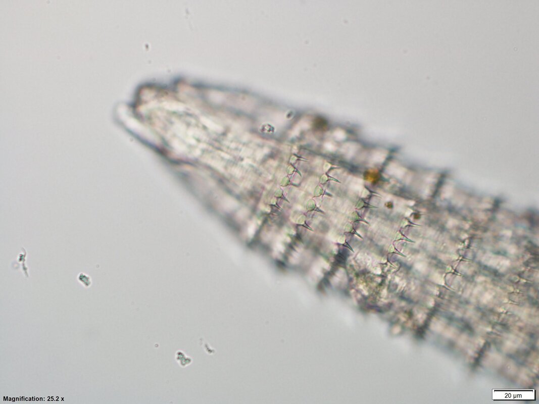
Image 3. You have goat to be kidding meA 1-month old Boer buckling was presented to the veterinarian for lethargy, recumbence, distended abdomen, tachycardia, and hypoglycemia. He was diagnosed with metabolic acidosis and was unable to recover. During necropsy several of the following specimens were recovered from hair. Video 1: Specimens recovered from the hair. Linognathus africanus (African goat louse) are Anoplurans or bloodsucking lice, with piercing mouthparts used for feeding. They spend their time on the hairs of the host, where they deposit eggs. All life stages occur on the host. Linognathus africanus and L. stenopsis, are species that can infest goats. Linognathus spp. first pair of legs is shorter than the second and third pairs, and L. africanus have a greatly expanded head behind the antennae compared to L. stenopsis. The African goat louse is distributed world-wide. Infestations by these lice are usually localized around the head, while the goat sucking louse (L. stenopsis) can be dispersed over the entire body. Lice are usually a nuisance, but in some cases severe infestations can cause scabby, and bleeding areas that may lead to bacterial infections. |

