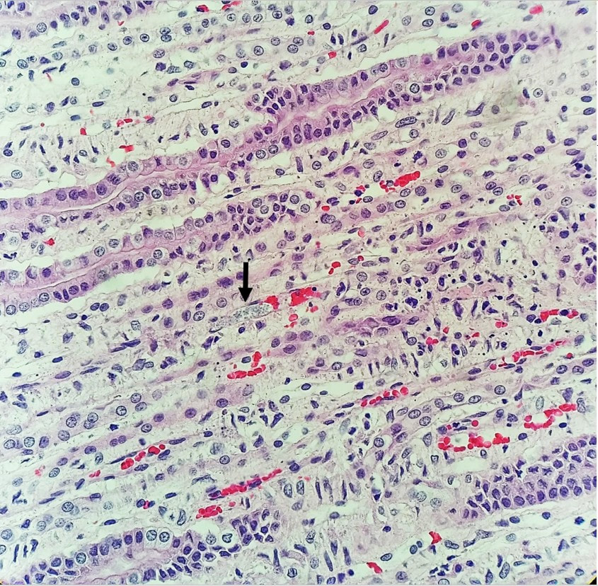
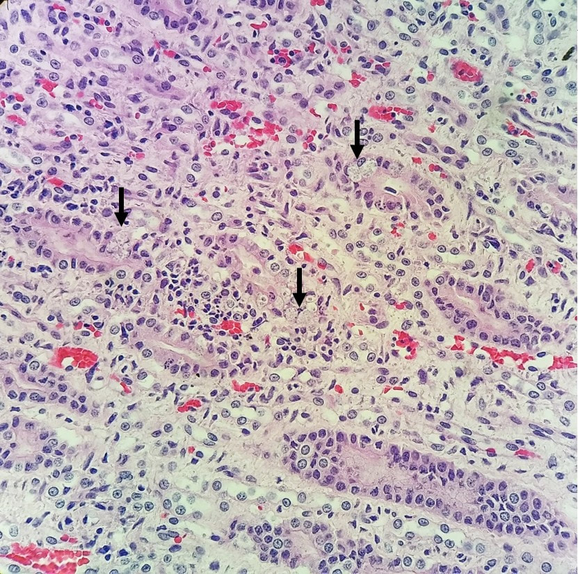
Hop to it!An approximately 6-year-old, intact female, mixed breed rabbit was euthanized after battling with the last stage of uterine adenocarcinoma and brought to necropsy. Besides confirming the adenocarcinoma, gross examination revealed some focal, irregular, depressed areas on the surface of the kidneys. On histologic examination, several spores were observed in the epithelial cells of the kidneys (Figures 1 & 2). Figure 1: Spores indicated by arrow. Figure 2: Spores indicated by arrows. Encephalitozoon cuniculi Encephalitozoon cuniculi is a spore-forming pathogen with worldwide distribution and has been isolated from various mammal species such as rabbits, shrews, mice, hamsters, muskrats, guinea pigs, goats, sheep, pigs, horses, domestic dogs, domestic cats, foxes, non-human primates, and man. Historically this pathogen was considered a protozoan parasite; however, due to its unique features, it is currently classified in the phylum Microsporidia. Infection occurs by ingestion or inhalation of spores shed in the feces, mucus, and urine of infected animals. Transplacental transmission has been also reported. Encephalitozoon cuniculi can cause granulomatous lesions in a wide range of tissues, but primarily affects the brain, kidney, or eyes. Most immunocompetent rabbits do not show any clinical signs; however, in some severe cases, granulomatous lesions associated with E. cuniculi can cause vestibular disease, chronic renal failure, lens rupture, pyogranulomatous uveitis, and cataracts. Ante-mortem diagnosis of E. cuniculi infection is challenging, and the gold standard is post-mortem histologic examination with immunochemical staining. In this case, the E. cuniculi infection was an incidental finding. References Molly Varga, 2014. Textbook of Rabbit Medicine, 2nd ed. Frank Kunzel, et al. 2018. Clinical signs, diagnosis, and treatment of Encephalitozoon cuniculi infection in rabbit. Veterinary Clinics of North America: Exotic Animal Practice. https://www.sciencedirect.com/science/article/pii/S1094919417302025?via%3Dihub#fig3 |
Archives
July 2024
Have feedback on the cases or a special case you would like to share? Please email us ([email protected]). We will appropriately credit all submittors for any cases and photos provided.
|


