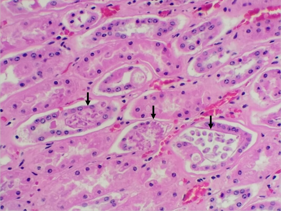
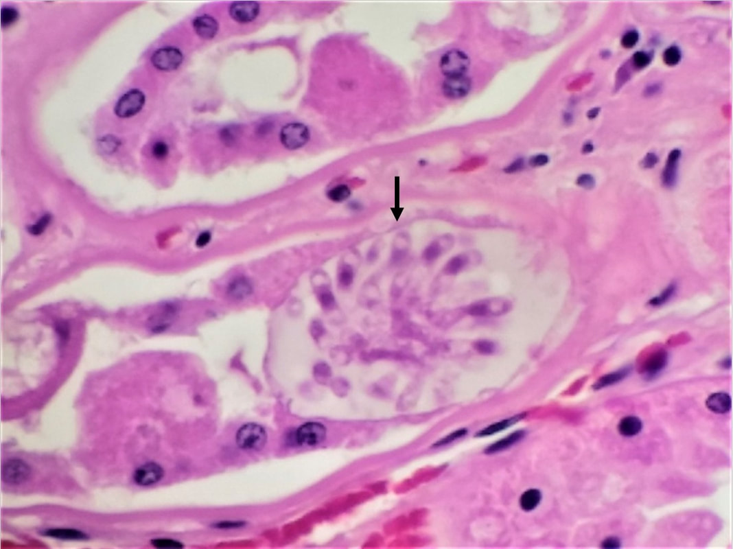
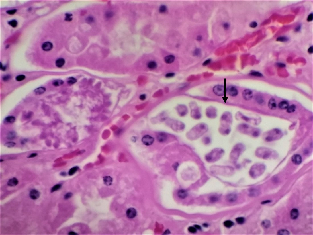
Incidental finding or cause of death? An approximately 8-year-old miniature donkey mare had difficulty foaling. The referring veterinarian provided aggressive supportive therapy but unfortunately the foal and mare did not survive. To determine the cause of death both carcasses were submitted for necropsy. Grossly, the mare was remarkably emaciated, and there were mild, chronic, multifocal ulcers in the stomach. Histologic examination of the stomach confirmed moderate, chronic-active, multifocal ulcerative gastritis with bacterial infection. Additionally, histologic examination of the mare’s kidney revealed multiple, variable sized parasites in the renal tubular epithelium (Figures 1-3). No abnormalities were detected grossly or microscopically in the foal. Figure 1: Various parasite stages in the renal epithelial cells (indicated by arrows). Figure 2: Multiple nuclei lie along the periphery – notice how they look like a flower, these are called “sporoblasts” (indicated by arrow). Figure 3: Free/Mature sporoblasts (indicated by arrow) – each of these sporoblasts undergoes further divisions to form sporocysts. Klossiella equi Klossiella equi is a protozoan parasite observed in the kidney of equines. The life cycle has not been fully understood; however, it is thought to be a direct life cycle. Sporocysts are passed in the urine and infection takes place by ingestion of sporulated sporocysts. Klossiella equi infection is thought to be fairly common throughout the world but rarely seen. Klossiella eqi is considered non-pathogenic and usually is not associated with clinical signs. In this case, K. equi infection was an incidental finding, and bacterial translocation from the gastric ulcers and subsequent septicemia was likely related to the death of the mare and foal. Special thanks to Dr. Rory Chia-Ching Chien, DVM, MSc, Anatomic Pathology Resident at Oklahoma State University, Center for Veterinary Health Sciences for sharing his case and photos! References: Taylor MA, Coop RL, Wall RL. Veterinary Parasitology (4thedition). Gardiner CH, Fayer R. Dubey JP. An Atlas of protozoan parasites in animal tissues. Further reading: Ballweber LR, Dailey D, Landolt G. (2012). Klossiella equiInfection in an Immunosuppressed Horse: Evidence of Long-Term Infection. Case Reports in Veterinary Medicine. 2012, 4. doi:10.1155/2012/230398. Reinemeyer CR, Jacobs RM, Spurlock GN. (1983). A coccidial sporocyst in equine urine. J Am Vet Med Assoc.182(11). 1250–1251. |
Archives
July 2024
Have feedback on the cases or a special case you would like to share? Please email us ([email protected]). We will appropriately credit all submittors for any cases and photos provided.
|


