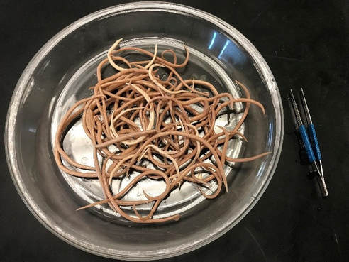
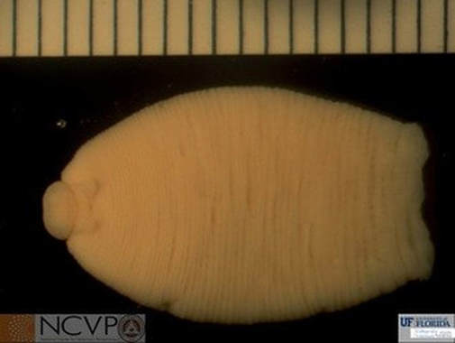
What a wasteAn 8-month-old male Thoroughbred horse presented to Oklahoma State University’s Equine Internal Medicine Service for suspected renal failure. Initial clinical signs including anorexia and weight loss were first observed 3 weeks prior, and the horse’s condition progressively worsened. The horse was humanely euthanitized and submitted for necropsy. Necropsy revealed large numbers of the following organisms: Many thanks to Erin Stayton, DVM and Resident in Anatomic Pathology at OSU-CVHS for providing this case. AnswerThese are adults of Parascaris (top photo) and Anoplocephala perfoliata (bottom photo). While it is unlikely that these parasites are responsible for the kidney failure in this animal, the intense infections may have contributed to overall ill-thrift. Parascaris are easily identified by their large, stout appearance and the presence of three prominent lips on the anterior end. The species designation for this common parasite of foals has been the subject of recent discussion; P. univalens appears more common than P. equorum in many areas. Once infected by ingestion of larvated eggs from the environment, Parascaris larvae migrate through the liver and lungs before returning to the small intestine as 4th stage larvae. Parascaris achieves patency in the small intestine between 75-80 days after infection. The migrating larvae are most often associated with pathogenicity, but in heavy infections, adults can cause colic due to intestinal impactions. Horses typically acquire immunity to these parasites by the second year of life. Anoplocephala perfoliata, a common tapeworm of horses, can be identified by the flattened body (8-25 cm in length) comprised of narrow proglottids, two pairs of suckers, and pronounced lappets on the anterior end. Infection is by ingestion of free-living oribatid mites containing cysticercoids from pasture. Within the horse, adult A. perfoliata can be found in the cecum, ileum, and clustered at the ileocecal junction. In light infections, clinical signs are most often absent. In heavy infections, gastrointestinal disturbances and unthriftiness may be observed. Mucosal ulceration in the area of A. perfoliata attachment is common, and inflammation at the ileocecal junction is linked to intestinal intussusception and colic. |
Archives
July 2024
Have feedback on the cases or a special case you would like to share? Please email us ([email protected]). We will appropriately credit all submittors for any cases and photos provided.
|


