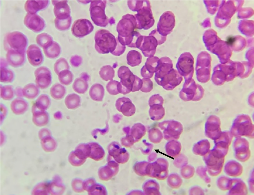
Puppy's parasite struggleA 14-week-old intact female Dalmatian, weighing 14 lbs (6.4 kg) was presented to a clinic in Gonzales, TX for dehydration and lethargy. Intake physical exam revealed ascites, pale mucous membranes, lethargy, and muffled lung sounds. Parvovirus antigen test was not detected in feces, but fecal centrifugal flotation demonstrated Ancylostoma sp. eggs 1+, packed cell volume was 20%, and total protein was 5.2. Patient died within 2 hours of presentation. Examination of stained thin blood films demonstrated the following (Image 1). Image 1: Stained blood film Case contribution: Alyssa Barta, DVM student, Oklahoma State University, Class of 2025, and Dr. John Withers. Pecan Grove Veterinary Clinic, Gonzales, TX. July 25, 2023. Trypanosoma cruzi trypomastigotes, observed in the blood of acutely infected dogs for a short time after infection. Morphological identification can be accomplished based on the presence of a terminal/subterminal kinetoplast that is darkly stained at the posterior end, a centrally located nucleus, an undulating membrane, and a flagellum that runs along the length of the trypomastigote body extending anteriorly beyond the margin of the flagellate. Trypomastigotes range in length from 12 to 30 µm and are often C-shaped (arrow) or J-shaped in fixed preparations. Despite finding trypomastigotes in this case, diagnosing Chagas disease in dogs is usually accomplished by T. cruzi antibody detection, using immunofluorescence antibody (IFA) tests in chronically infected dogs. Trypanosoma cruzi infected dogs are more likely to have ventricular arrhythmias, combinations of ECG abnormalities, and cardiac troponin I (cTNl) >0.129 ng/mL. Surveys of dogs in T. cruzi endemic areas, showed that infected dogs were significantly younger than negative dogs and the highest prevalence of infection were in non-sporting and toy breed dogs. Odds of T. cruzi infection were 13 times greater among dogs where there was an infected housemate or littermate. |
Archives
July 2024
Have feedback on the cases or a special case you would like to share? Please email us ([email protected]). We will appropriately credit all submittors for any cases and photos provided.
|

