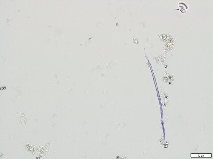
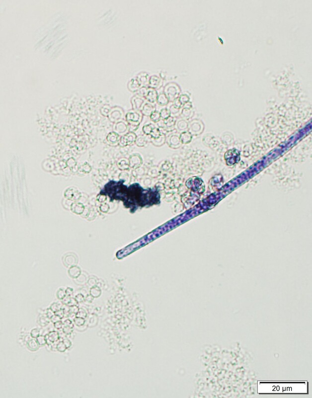
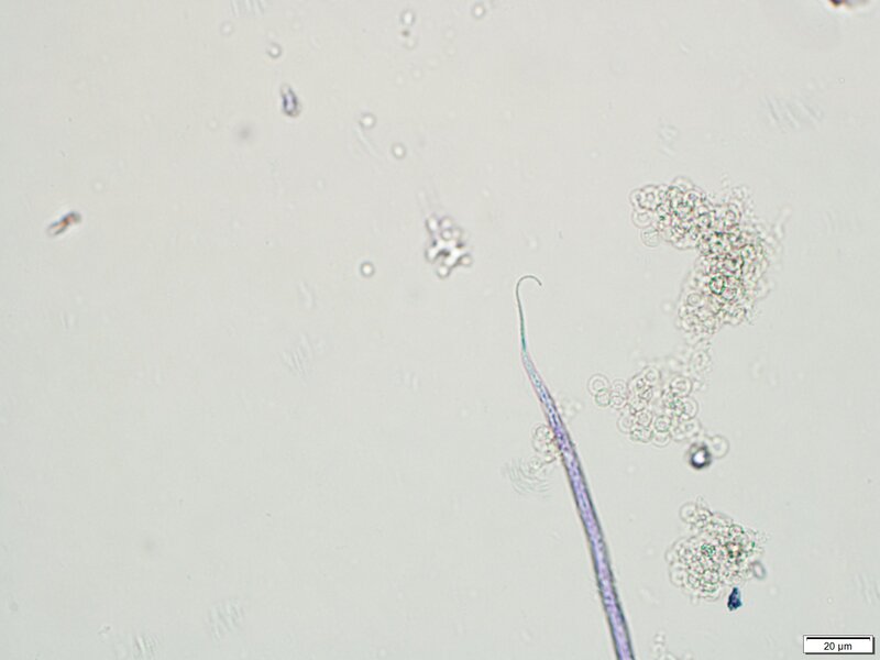
Dog"ged" microfilariaeA blood sample from a 21-month old female Boston Terrier Mix, was submitted to the parasitology diagnostic lab to be tested for Dirofilaria immitis detection. The dog had a history of microfilaremia in 2020 but no antigen was detected during that same year. In September 2021, during the annual wellness a snap test was performed at the clinic with no antigen detected, but microfilaria was again observed on the direct smear. To rule out the presence of antigen-antibody complexes, the serum sample was heat treated at the diagnostic lab, but no antigen was detected. The Modified Knott’s test showed the presence of the following microfilariae. (Images 1-3) Size average 275 µm in length x 5 µm in width.Images 1-3: Key features for identification. Acanthocheilonema reconditum, is a member of the subfamily Onchocercinae that infects canids. It is transmitted by fleas and the louse Heterodoxus spiniger. The adults are located in the subcutaneous tissues where they do not cause any pathology. The importance of this parasite lies on the confusion that its microfilariae can create on the diagnosis of Dirofilaria immitis infection. Both microfilariae are similar and can be easily confused, however morphological characteristics and motility can help differentiate these two microfilariae. Observing microfilariae in puppies and young dogs with no D. immitis antigen detection can be explained by infection of A. reconditum such as this case or transplacental transmission of D. immitis. |
Archives
July 2024
Have feedback on the cases or a special case you would like to share? Please email us ([email protected]). We will appropriately credit all submittors for any cases and photos provided.
|


