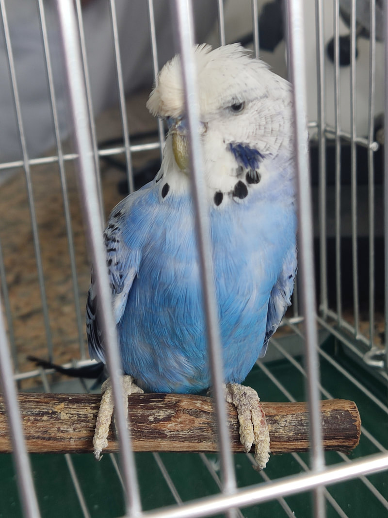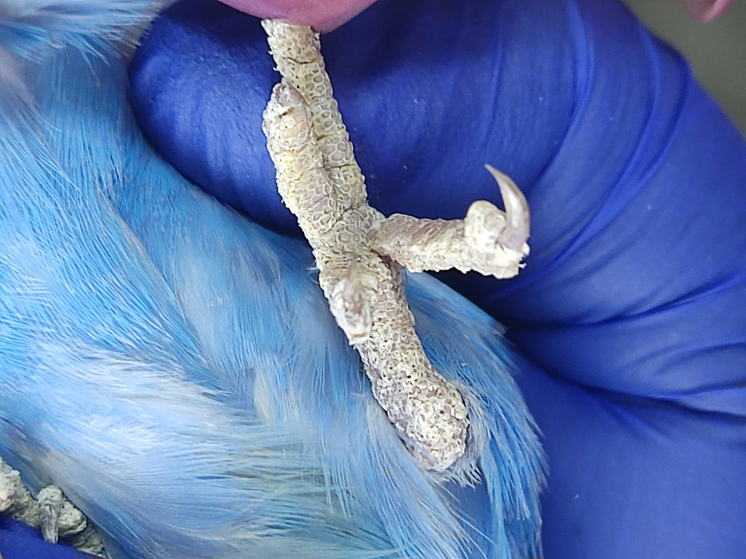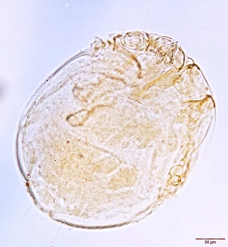
Hope for a New Year's ResolutionA parakeet pet was presented to the College of Veterinary Medicine Clinic, Southwestern University in Philippines, with localized thickened, crusty lesions on the legs and feet, and scales lifting off on the area. No signs of pruritus were noticed. (Images 1-3) Microscopic examination of loose leg scales revealed the following. (Image 4) Case contribution: Erlah Shermaine Roble, DVM, Assistant Professor at the College of Veterinary Medicine. Southwestern University PHINMA. Philippines Images 1-3: Gross lesions upon presentation Image 4: Microscopic finding of leg scales Knemidocoptes sp. These mites burrow in the epidermis of the legs on birds, causing scales to lift and become loosened and the legs to become thickened and deformed. K. pilae is the species that commonly infest parakeets while K. mutans and K. jamaiciensis infest gallinaceous birds and canaries respectively. This case was resolved after oral administration of Ivermectin in drinking water. Christmas SurpriseA 2-year-old female Mongolian gerbil was submitted for necropsy to determine the cause of death as the owner has had multiple losses in the household. During necropsy, six specimens collected from the proximal area of the small intestine were incidentally found. All of the specimens recovered were identified as females containing spindle shaped eggs that measured 115 µm to 140 µm by 30 µm to 60 µm. (Images 1-3). Case provided by: Tiana Sanders DVM, PhD Student, Texas A&M Image 1: Eggs, 10x Image 2: Caudal end, 10x Image 3: Anterior end, 10x Dentostomella translucida. Known as the “Gerbil Pinworm” is a threadworm found in the small intestine of gerbils. Females typically measure 1 cm to 3 cm in length and males are 0.6 cm to 1 cm. The identifying features of D. translucida include a mouth with no lips, five unequal teeth per esophageal sector, and a thick, translucent, transversely striated cuticle. Typically, gerbils infected with D. translucida are asymptomatic and diagnosis may be difficult, as these parasites do not deposit their eggs around the perianal region. Worms on the BrainAn adult female woodchuck presented to a wildlife clinic in New York state after being found on the side of the road. The woodchuck had multifocal crusts and pustules over most of the body, with skin sloughing, with mites and fleas noted on external examination. The animal displayed neurologic signs, with, minimal response to handling, and paddling reported. A dewormer and antibiotics were administered, but despite treatment, the woodchuck continued to decline, with no interest in food by the third day. Euthanasia was elected, and the animal was submitted for necropsy following a negative rabies test. Histologic examination of the brain revealed the following: Case contribution: Timothy Wu, MS, DVM, Dipl. ACVP Image 1 and 2: Histologic findings in the brain Ascarid larva migrans (likely Baylisascaris sp.) Histologic evaluation reveals numerous up to 60 um diameter nematodes, determined to be the larval stage, given the absence of genital tracts within the examined sections. The presence of lateral alae (arrow), coelomyarian-polymyarian musculature, and a uninucleate intestine are consistent with ascarid larvae. While other ascarids, such as Toxocara canis cannot be ruled out, the most likely diagnosis in this case is considered to be Baylisascaris sp. There are many species of Baylisascaris affecting wildlife, including Baylisascaris columnaris in skunks, Baylisascaris transfuga in bears, and Baylisascaris laevis in woodchucks. The most commonly discussed species is Baylisascaris procyonis, which affects raccoons, and can cause visceral larva migrans in a wide range of hosts, including humans. Ascarid migration of the central nervous system, as in this case, can cause severe clinical signs. Although quick diagnosis and treatment with corticosteroids and albendazole can prove effective, this is not always plausible, as diagnosis can prove difficult. Hidden sidekickA 7-year-old male Siberian husky was taken to a clinic for a wellness check. The dog lives in a suburban area in a house with close access to a wooden area. According to the owner, he was acting playful and eating normal. The gross fecal exam was normal and the centrifugation with sugar solution revealed the following: Image 1 and 2: Findings of centrifugation fecal with sugar Sarcocystis sp. oocysts. Dogs and other carnivores are definitive hosts of Sarcocystis sp. while ruminants, horses, swine and rabbits can act as intermediate hosts. Fully sporulated oocysts are eliminated thorough the dog’s feces and the schizogony and sarcocysts formation occurs in the intermediate hosts. Definitive hosts usually do not display clinical signs but the schizogony in the endothelium of the herbivore may result in serious disease. A known pathogenic species for equids is Sarcocystis neurona with opossums acting as definitive hosts. Now serving eggs a la parasiteA backyard flock owner in Minnesota found the following specimen when she cracked one of their eggs. Contributors: Lindsey King DVM class 2024 at OSU Kellee Sundstrom, MS, Veterinary Pathobiology, OSU Prosthogonimus macrorchis, are poultry flukes found within the oviduct. Birds are infected with P. macrorchis after consuming an infected dragonfly. Because of the use of aquatic invertebrates as intermediate hosts, this parasite is commonly found in fowl surrounding water features, especially the Great Lake area. Potential clinical signs are weight loss, malformed eggs, or decreased egg production. Adult flukes are small, approximately 8mm long, often found within the oviduct on necropsy. A recent case report also described P. macrorchis in the egg of a backyard chicken. At this time, there is no labeled dewormer for P. macrorchis in poultry. Puppy's parasite struggleA 14-week-old intact female Dalmatian, weighing 14 lbs (6.4 kg) was presented to a clinic in Gonzales, TX for dehydration and lethargy. Intake physical exam revealed ascites, pale mucous membranes, lethargy, and muffled lung sounds. Parvovirus antigen test was not detected in feces, but fecal centrifugal flotation demonstrated Ancylostoma sp. eggs 1+, packed cell volume was 20%, and total protein was 5.2. Patient died within 2 hours of presentation. Examination of stained thin blood films demonstrated the following (Image 1).  Image 1: Stained blood film Case contribution: Alyssa Barta, DVM student, Oklahoma State University, Class of 2025, and Dr. John Withers. Pecan Grove Veterinary Clinic, Gonzales, TX. July 25, 2023. Trypanosoma cruzi trypomastigotes, observed in the blood of acutely infected dogs for a short time after infection. Morphological identification can be accomplished based on the presence of a terminal/subterminal kinetoplast that is darkly stained at the posterior end, a centrally located nucleus, an undulating membrane, and a flagellum that runs along the length of the trypomastigote body extending anteriorly beyond the margin of the flagellate. Trypomastigotes range in length from 12 to 30 µm and are often C-shaped (arrow) or J-shaped in fixed preparations. Despite finding trypomastigotes in this case, diagnosing Chagas disease in dogs is usually accomplished by T. cruzi antibody detection, using immunofluorescence antibody (IFA) tests in chronically infected dogs. Trypanosoma cruzi infected dogs are more likely to have ventricular arrhythmias, combinations of ECG abnormalities, and cardiac troponin I (cTNl) >0.129 ng/mL. Surveys of dogs in T. cruzi endemic areas, showed that infected dogs were significantly younger than negative dogs and the highest prevalence of infection were in non-sporting and toy breed dogs. Odds of T. cruzi infection were 13 times greater among dogs where there was an infected housemate or littermate. |