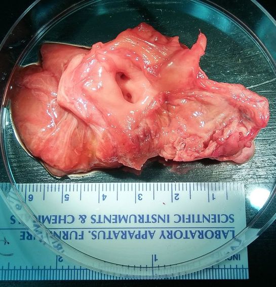
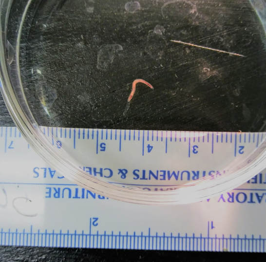
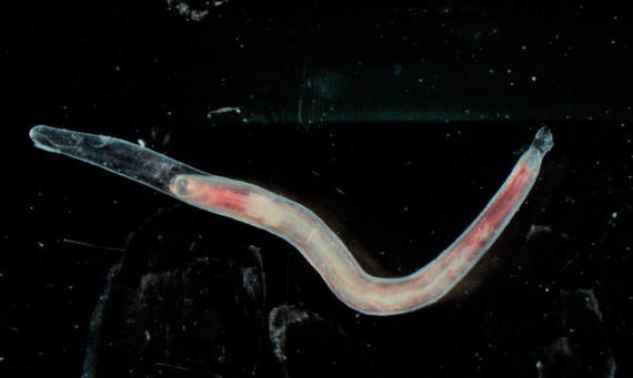
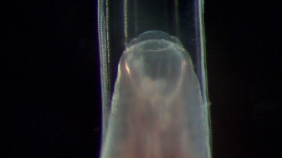
What a Rare Find!!An emaciated horse with no known previous history was rescued and presented to Large Animal Services at Oklahoma State University. The horse was humanely euthanized and sent to necropsy the next day. On necropsy, a pathologist found the severe local arteritis, especially in the cranial mesenteric artery, with a worm in the lesion. The cranial mesenteric artery. The wall was very thickened and fibrous. A nematode worm was also detected in the lesion of the cranial mesenteric artery. A nematode worm detected in the cranial mesenteric artery of the emacinated horse. The cranial end of the nematode. Note there was a big buccal cavity and 2 teeth in the bottom. Special thanks to Dr. Kamilyah R. Miller at College of Veterinary Medicine, Tuskegee University & Dr. Yoko Nagamori at Center for Veterinary Health Sciences, Oklahoma State University. AnswerStrongylus vulgaris -- This is a sheathed Strongylus vulgaris L5. There were 2 teeth in the bottom of the buccal cavity, which distinguish it from other Strongylus species. Migrating S. vulgaris larvae can cause thickening of the cranial mesenteric artery, which impairs intestinal blood flow and leads to colic. Clinical signs include fever, anorexia, weight loss and depression. This case was a rare find of a fourth stage larvae molting into an adult Strongylus vulgaris worm. An infected animal sheds eggs, and under the right humidity and temperature the eggs develop into L3. A naïve animal ingests the L3 while grazing, the L3 penetrate the submucosa of the cecum and ventral colon, molt into L4 and penetrate smaller arterioles and migrate through the intima of the vessels to the larges branches of the cranial mesenteric artery. As the larvae grow larger, and occlude smaller vessels, the larvae travel to the cranial mesenteric artery and become encapsulated in nodules where they molt to immature adults. Some larvae are found in the arteries, before the fifth molt occurs, with the sheath still present. The immature adults travel back to the cecum and colon, mature and reproduce producing eggs 6 months after the animal becomes infected. Comments are closed.
|
Archives
July 2024
Have feedback on the cases or a special case you would like to share? Please email us ([email protected]). We will appropriately credit all submittors for any cases and photos provided.
|


