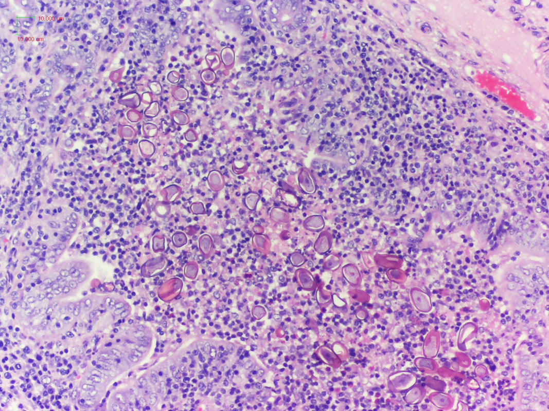
Unlucky rabbitsA juvenile female rabbit was presented for necropsy at the Veterinary Medical Diagnostic Laboratory, University of Missouri. The rabbit was part of an ongoing case of about 200 rabbits displaying various clinical signs including diarrhea, and observation of Eimeria spp. oocysts during fecal examination. At necropsy, white foci (1-3 mm diameter) on the liver and kidney were observed grossly. The following was observed during histological examination of the liver: Image 1: Histological liver preparation. Thanks to Dr. Alexis Carpenter, DVM and pathology resident at the University of Missouri, for providing the histological image and anamnesis of the case. Hepatic coccidiosis, caused by Eimeria stiedae. Some rabbits are asymptomatic; however, the disease can be fatal, especially in young rabbits. Heavily infected rabbits show signs related to decreased hepatic function and bile duct obstruction. Anorexia, debilitation and diarrhea or constipation can occur. The abdomen might be enlarged, and the animal may be icteric. Infection results from ingesting sporulated oocysts that undergo excystation in the duodenum. Enlargement of the liver, gall bladder, and bile duct may be seen at necropsy with multi-focal nodules caused by hyperplasia and cystic enlargement of the bile duct epithelium. Various life cycle stages of the organism may be seen histopathologically with oocysts in bile duct lumina. Comments are closed.
|
Archives
July 2024
Have feedback on the cases or a special case you would like to share? Please email us ([email protected]). We will appropriately credit all submittors for any cases and photos provided.
|

