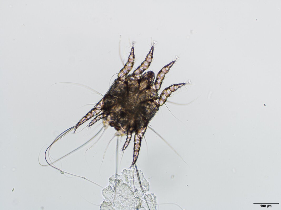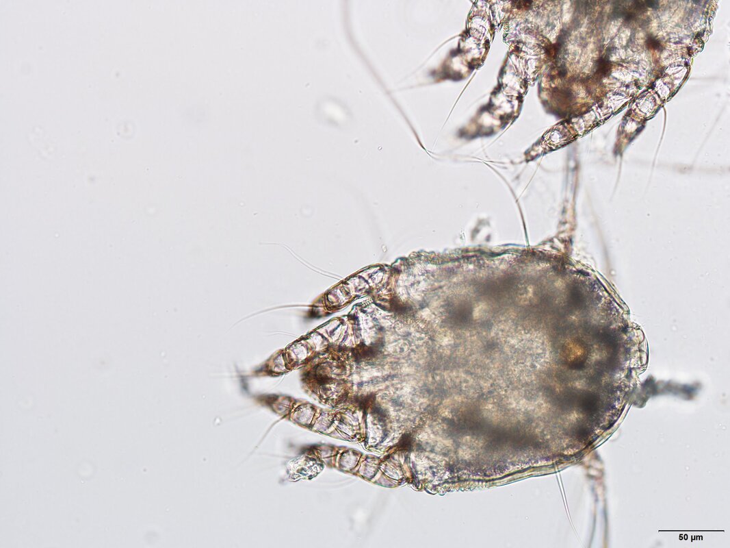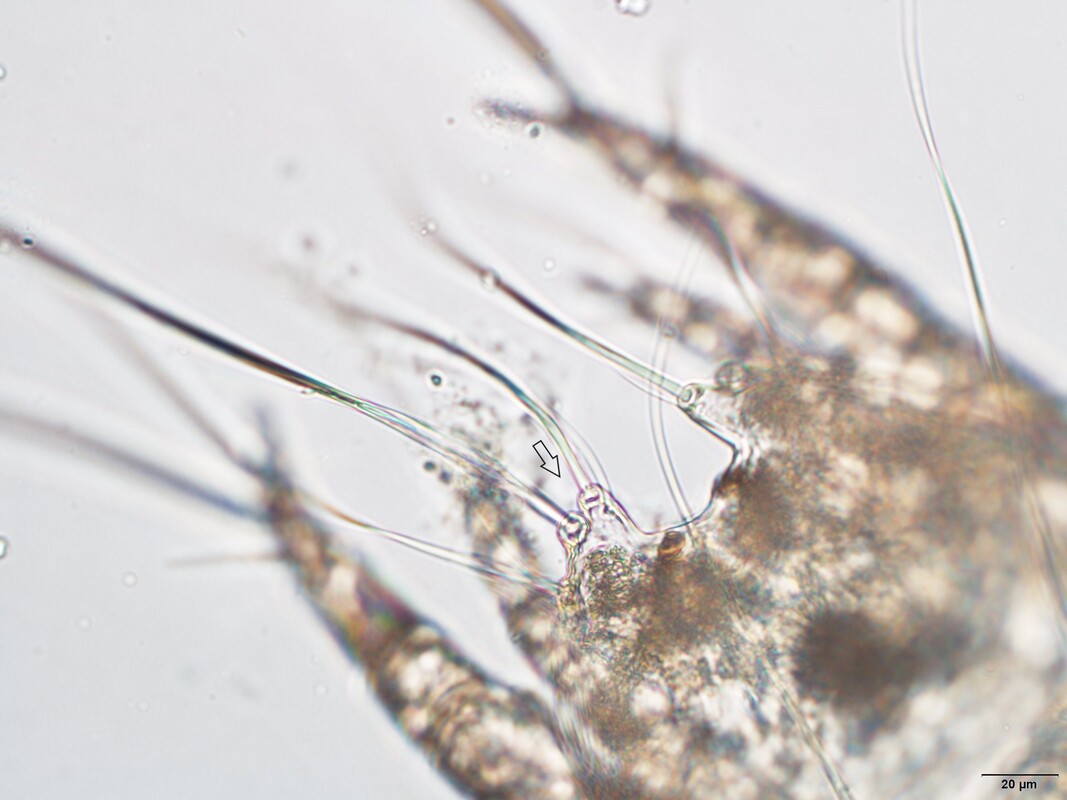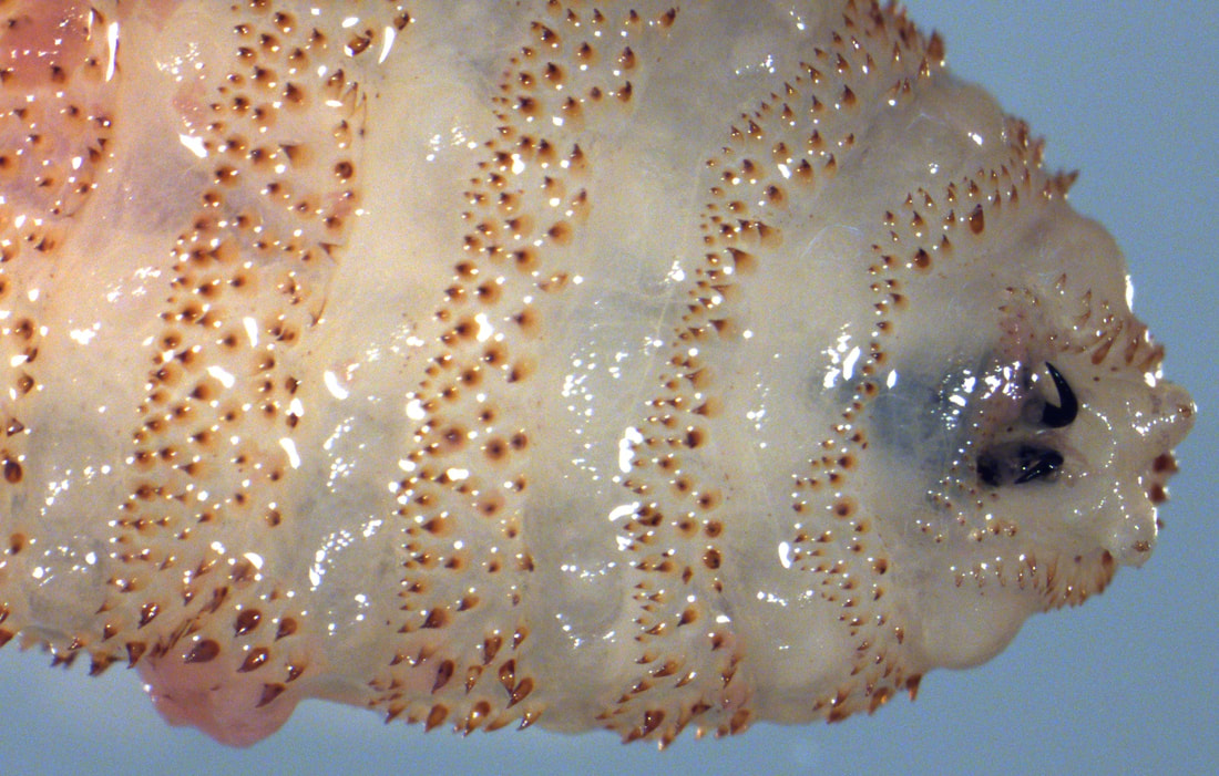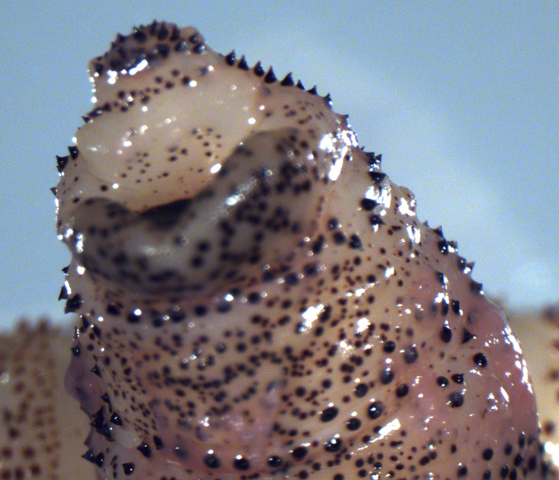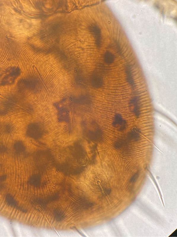
Indiana Newcomer!A 3-year-old, male, intact domestic shorthair cat showed signs of anorexia, vomiting, drooling, tachypnea, ataxia, and twitching. The cat tested positive for Feline Infectious Peritonitis. He did not respond to treatment and died due to Cardiopulmonary arrest (CPA). Necropsy was performed at the Indiana Animal Disease Diagnostic Laboratory, Purdue University. This parasite egg shown in the image was an incidental finding on centrifugal fecal flotation performed using Sheather’s solution. 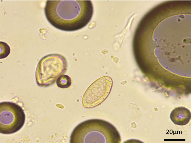 Image 1: Ova found on fecal examination (400x magnification) This month’s case was a contribution of Dr. Vishnu Manikantan, Purdue University. Opisthorchids are trematodes residing in the gall bladder and bile duct of many vertebrates. The eggs (22-35µm X 11-19 µm) are thin-shelled and operculated with a yellowish-brown color. The presence of a prominent lip around the rim of the operculum and the adjacent shell is a distinguishing feature of these eggs. In cats, Metorchis conjunctus, M. albidus, and Amphimerus pseudofelineus are the Opisthorchids reported from parts of the United States and Canada. Parasites belonging to the family Opisthorchiidae have an indirect life cycle involving two intermediate hosts- mollusks and freshwater fish. Opisthorchid infection is not known to create severe health effects in the definitive host. Nevertheless, there have been occasional reports of clinically significant liver damage. Eggs can be recovered using centrifugal fecal flotation, but sedimentation tests are preferred. The "mite"mare before ChristmasA 2-year-old African Pygmy Hedgehog, was presented with history of mucoid diarrhea for the past 24 hours. A centrifugal fecal flotation was performed and several of the following mites were observed. Image 1 & 2: Mites found on fecal flotation Image 3: Arrow pointing to trilobate laminate projection Caparinia tripilis are Psoroptid mites that infest hedgehogs as well as other mammals. The mites feed on skin cells and epidermal debris. Two species are known to infest hedgehogs, C. tripilis and C. erinacei, with the first one being more pathogenic and forming clusters on its host. Caparinia tripilis has a posterior end of the abdomen with trilobate laminate projection on either side and contain a long seta (Image 3) while C. erinacei has two bilobate laminate projections. The mites burrow into the skin of ears, head, and sides of the legs causing irritation, pruritus, and self-trauma that can lead to infection. On this case, the mucoid diarrhea was not caused by the mite infestation. Oh deer!A white-tail deer from a Northeast Oklahoma farm, was submitted for necropsy to the Oklahoma Diagnostic Laboratory during the month of August 2022. During necropsy the following larva was found on the esophagus. Images 1-3: Larva found in the esophagus Images 4-5: Posterior spiracles of recovered larva Hypoderma lineatum, also called heel flies or gadflies. The adult flies deposit eggs on the hair of cattle, deer and sometimes horses. The eggs hatch within a week and migrate through the connective tissues of the host. The larva of H. lineatum accumulate in the esophagus during migration and remain there for about 3 months. After, they migrate to the subcutaneous tissues of the back, forming lumps or warbles and cutting breathing holes in the skin of the host. When the larva emerges from the warble they measure approximately 25 mm long with a light brown color. Gerbil with an itchA Bushy Tailed Jird/Gerbil was presented to Texas A&M college of veterinary medicine with a two-week history of progressive pruritus on the left ear. The pruritus progressed to the point where the external ear was scabbed and scratched till it was raw (Image 1). The gerbil was not on any medication or prevention and no other animals in the same cage appeared affected. Several specimens, as the one observed on the following images were collected from the gerbil (Image 2-3). Image 1: Lesion on ear of gerbil Image 2-3: Specimens collected from gerbil This month’s case was a contribution of Tiana Sanders and Dr. Gui Verocai. Ornithonyssus bacoti. Known as the “tropical rate mite” is a bloodsucking mesostigmatid mite. They can be distinguished from Liponyssoides sanguineus, another mite that typically parasitizes rodents, by observing an anterior anus within the anal plate compared to a centrally located anus in Liponyssoides. Liponyssoides also has long whip-like chelicerae whereas Ornithonyssus have well-developed chelicerae that are the same in diameter throughout. Ornithonyssus is zoonotic and results in “Tropical Rat Dermatitis” in humans. It also harbors other pathogenic agents such as Francisella tularensis, Yersenia pestis, Borrelia burgdorferi and Rickettsia typhi. Annoying companionThe following parasites were recovered from the large intestine of a colony of prairie voles: Image 1-3: Key features for identification. Image 4: Egg image. Syphacia obvelata female. This parasite is a nematode of the order Oxyurida commonly found in mice and gerbils. Eggs can be detected by routine flotation techniques or found on the skin in the perineal region of infected animals. Infections are usually subclinical. For goat's sake!A herd of goats, from central Oklahoma submitted fecal samples to the Oklahoma Diagnostic Parasitology lab for fecal egg counts (FEC) and Trichostrongyles larval culture. Fecal samples look normal in appearance with FEC ranging between 500 to 2000 EPG. During larval identification the following parasites were observed:  Image 1: First stage larvae identified in culture.  Image 2: Tail of first stage larvae with dorsal spine. Muellerius capillaris This parasite is usually nonpathogenic, except when heavy infections occur. It is a Metastrongylid lungworm that infects sheep and goats, with adults living in the lung tissue or reactive nodules in the lungs. The first stage larvae are detected antemortem by the Baermann technique and characterized by having a tail with a dorsal spine. |