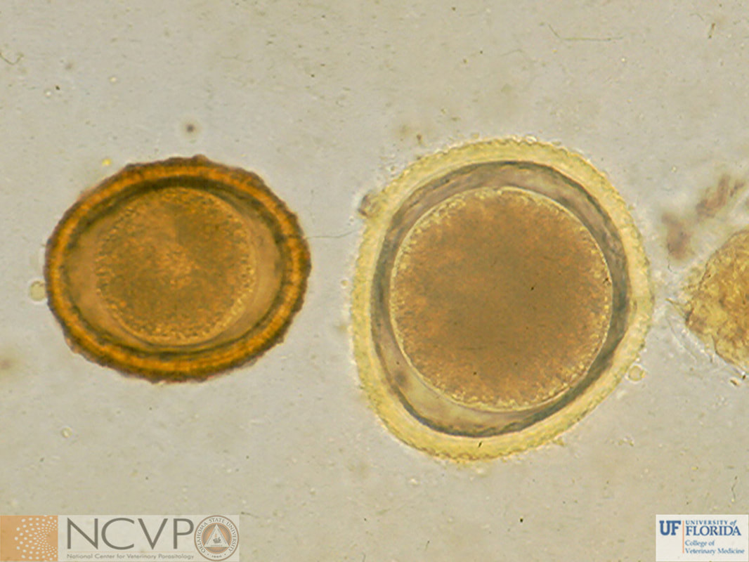
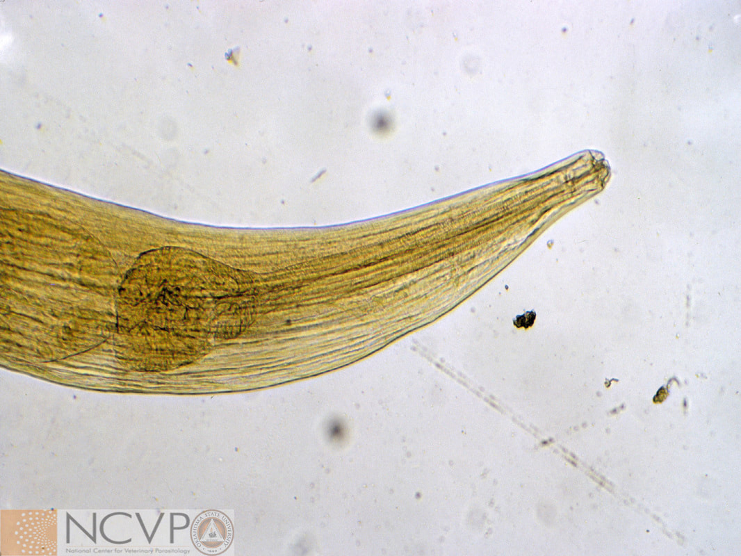
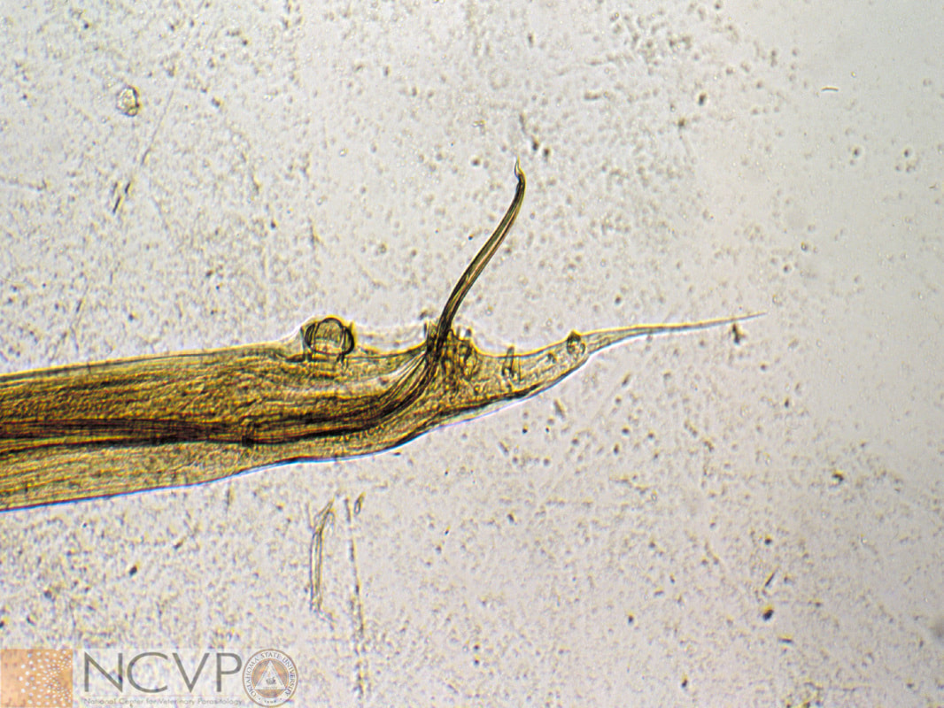
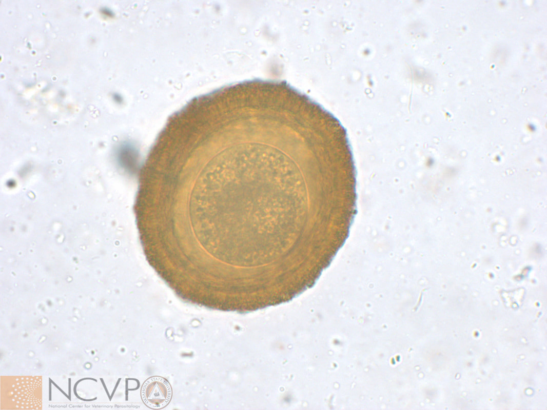
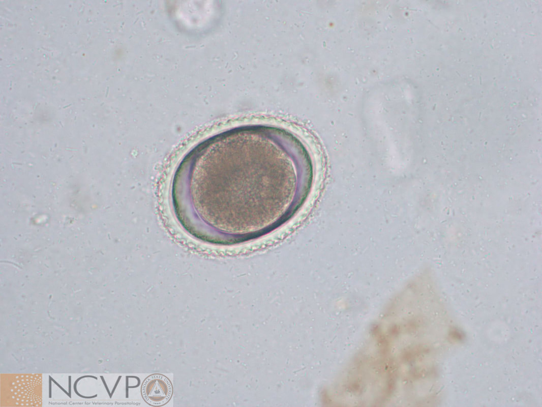
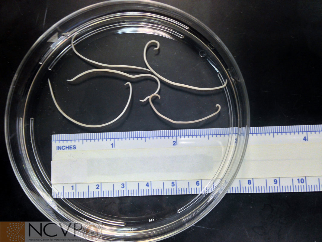
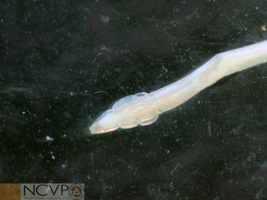
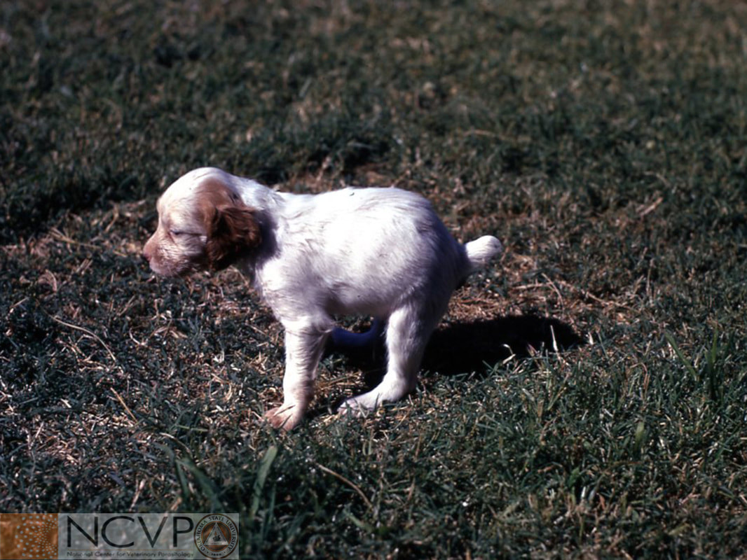
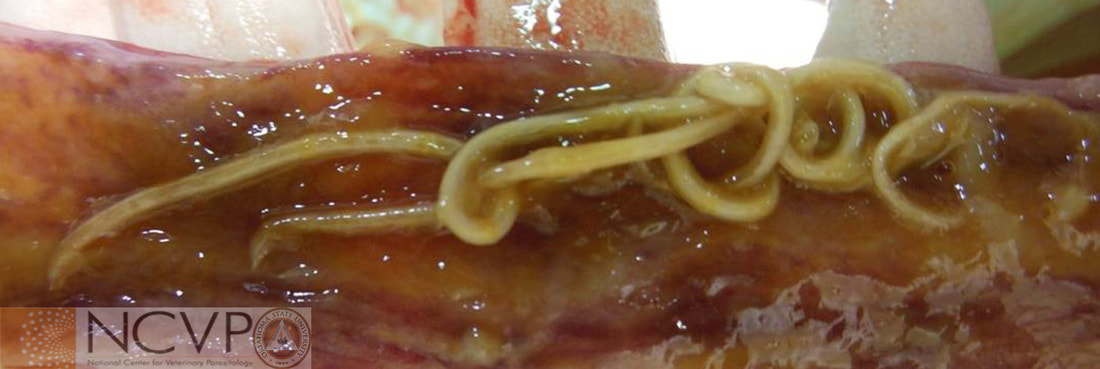
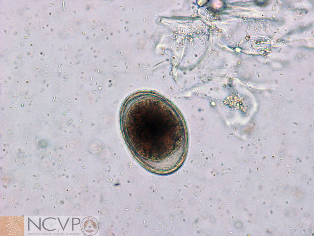
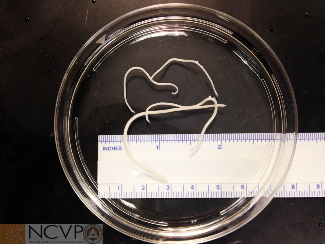
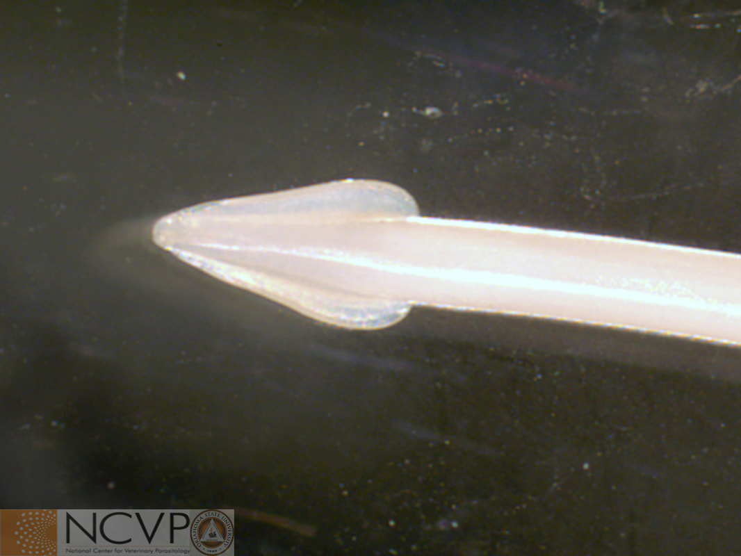
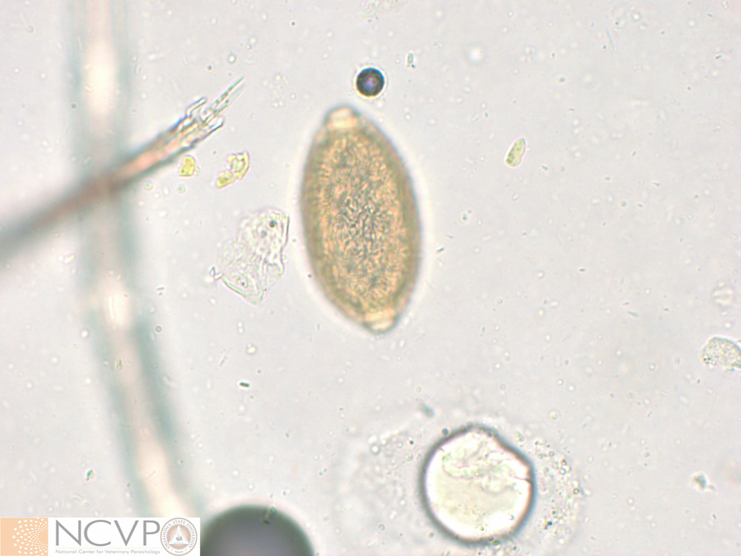
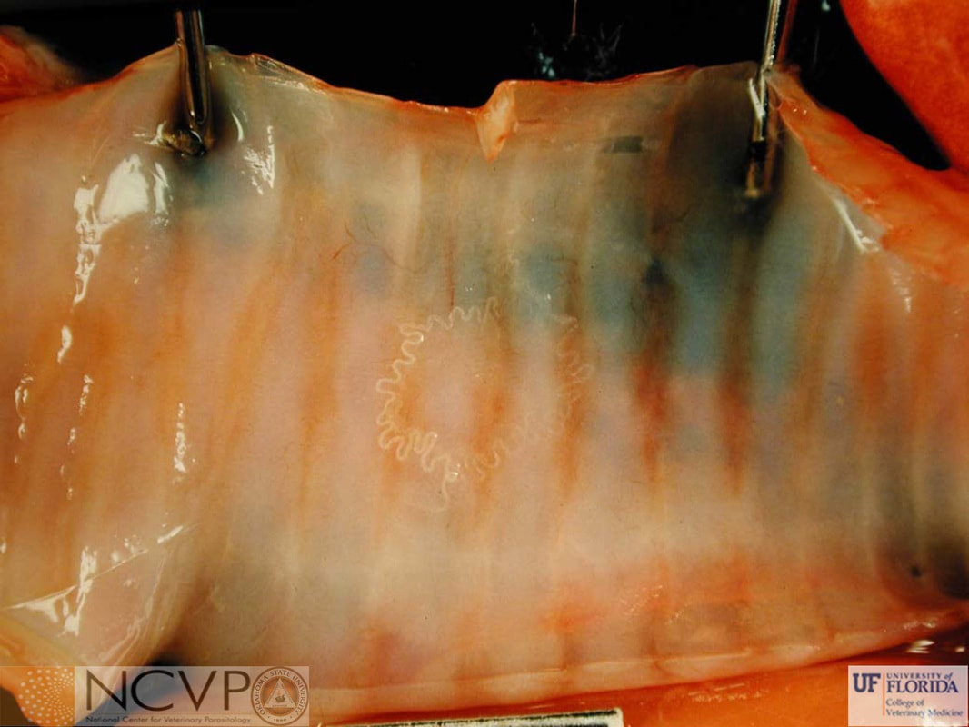
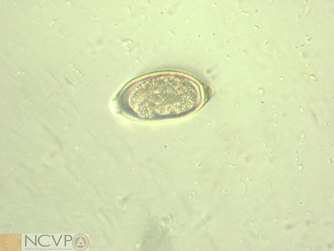
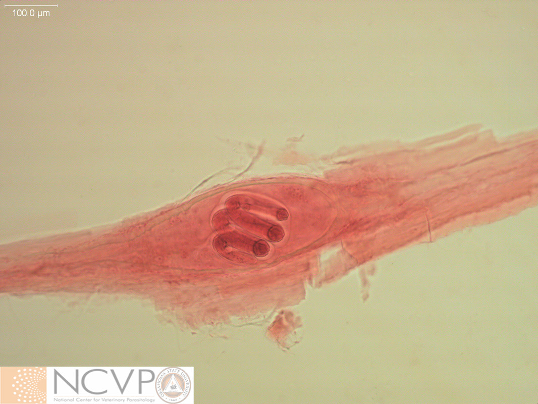
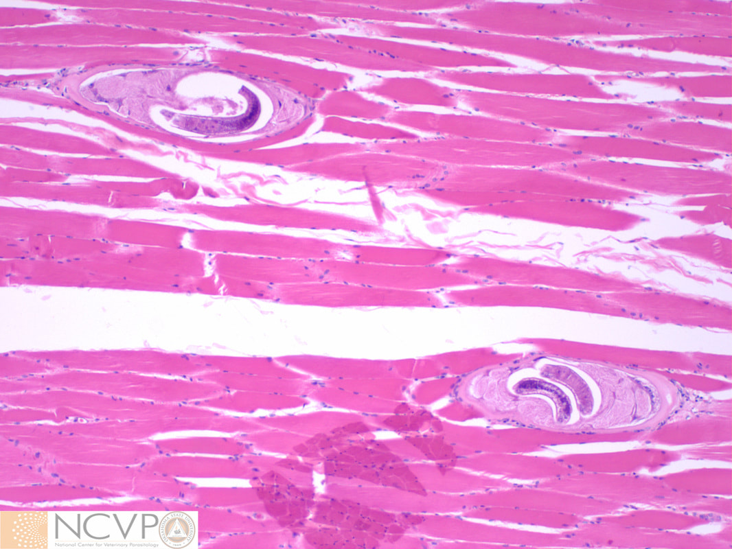
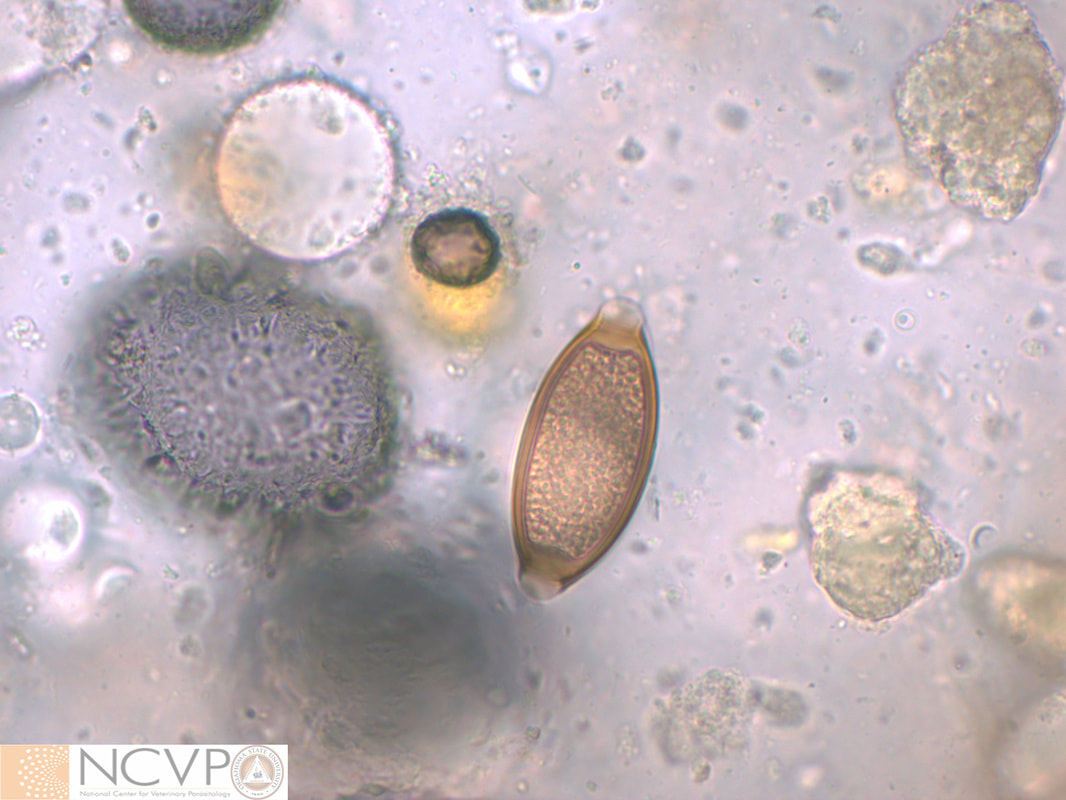
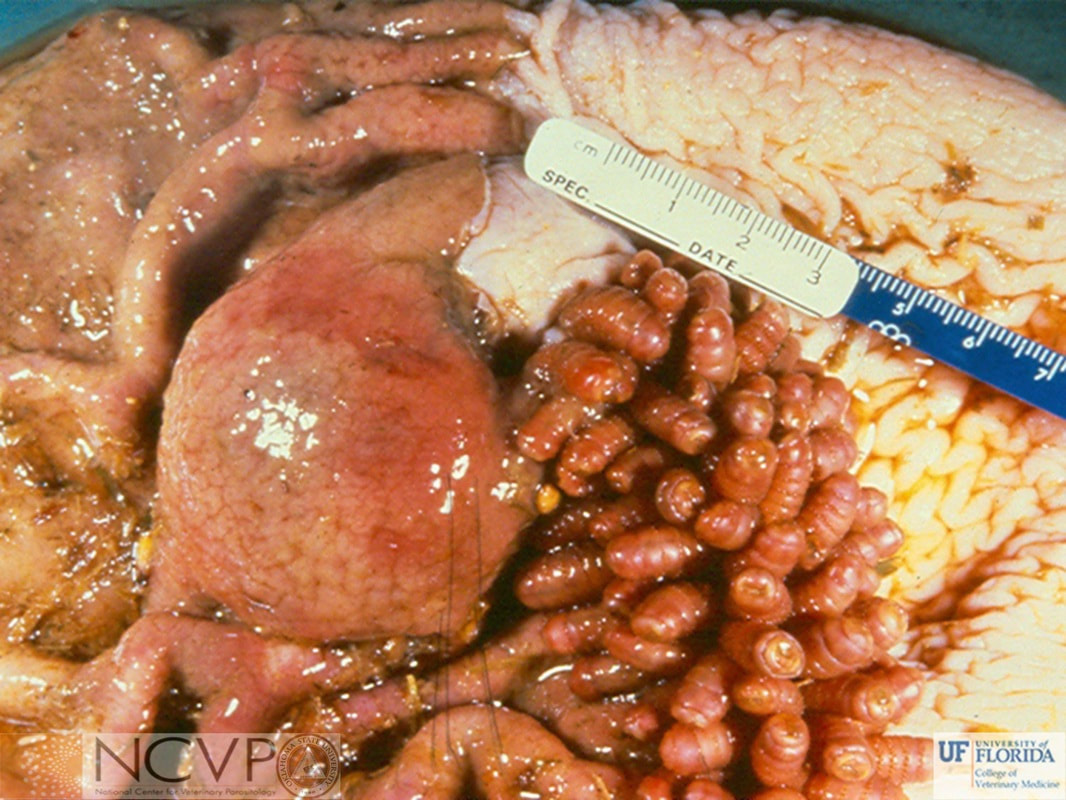
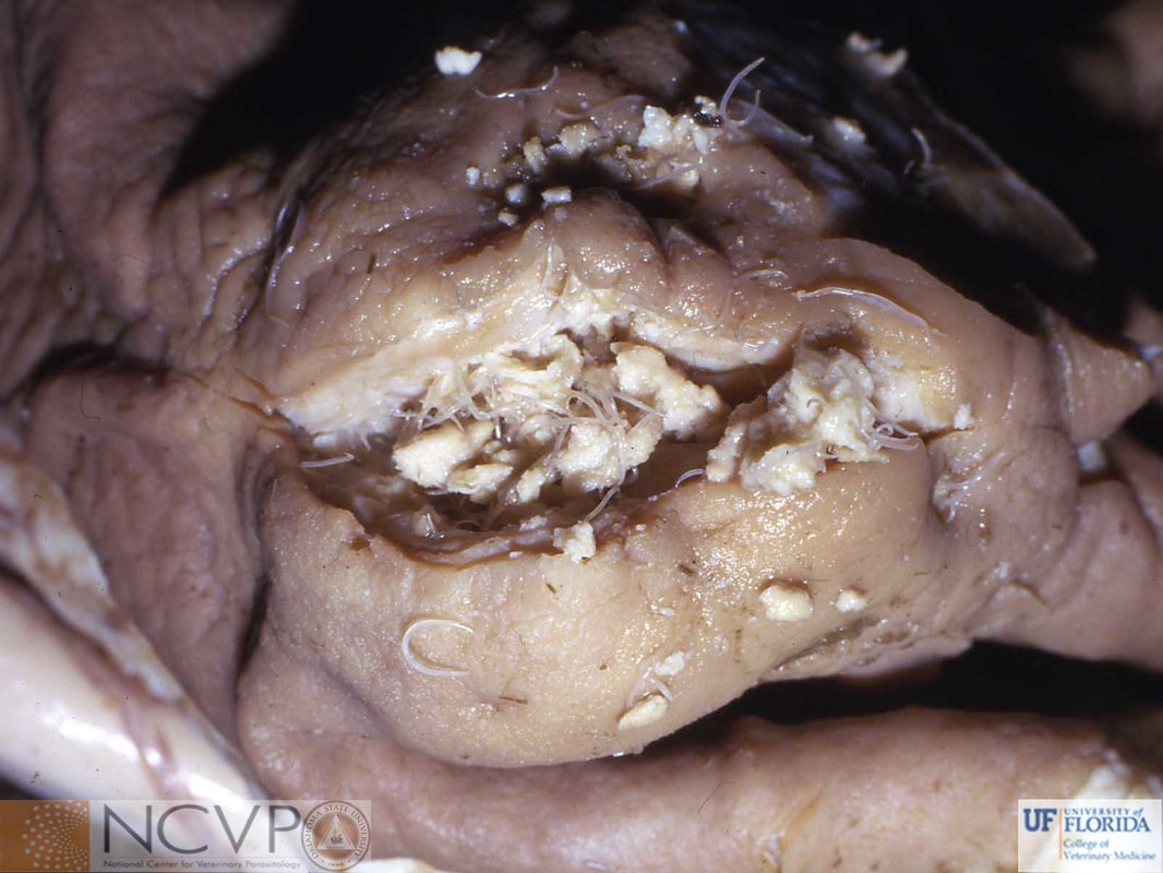
Ascaridoidea - Ascarid Roundworms CruziidaeFilaroideaOxyuroidea - PinwormsTrichinelloidea - Whipworms, Capillarids, and Trichinella spp.SpiruroideaRhabditoidea |
Categories |

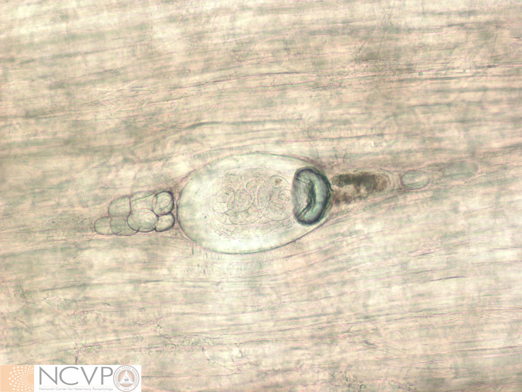
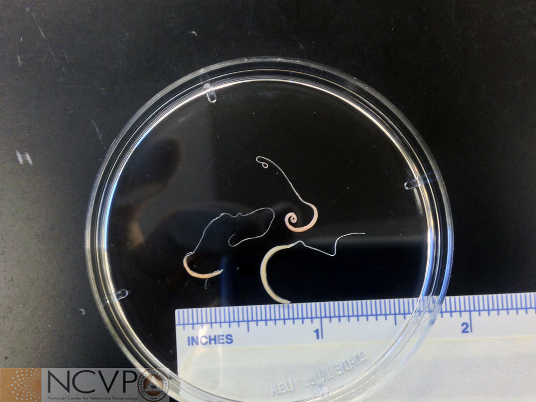
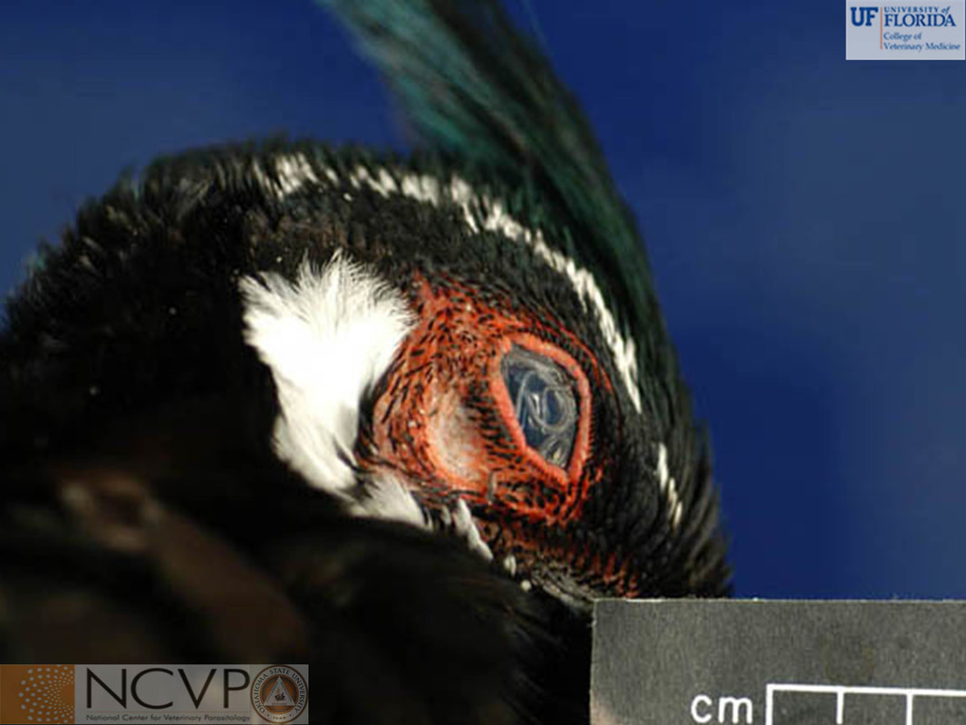
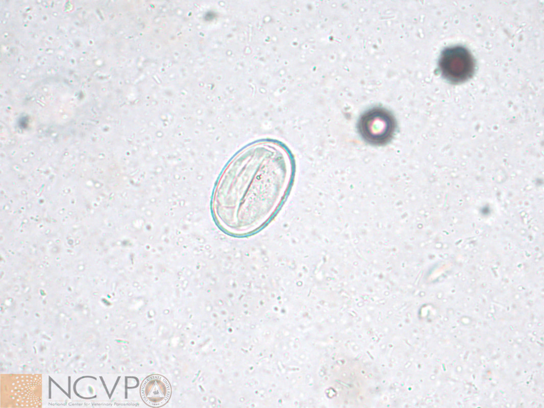
Ascaridoidea - Ascarid Roundworms CruziidaeFilaroideaOxyuroidea - PinwormsTrichinelloidea - Whipworms, Capillarids, and Trichinella spp.SpiruroideaRhabditoidea |
Categories |