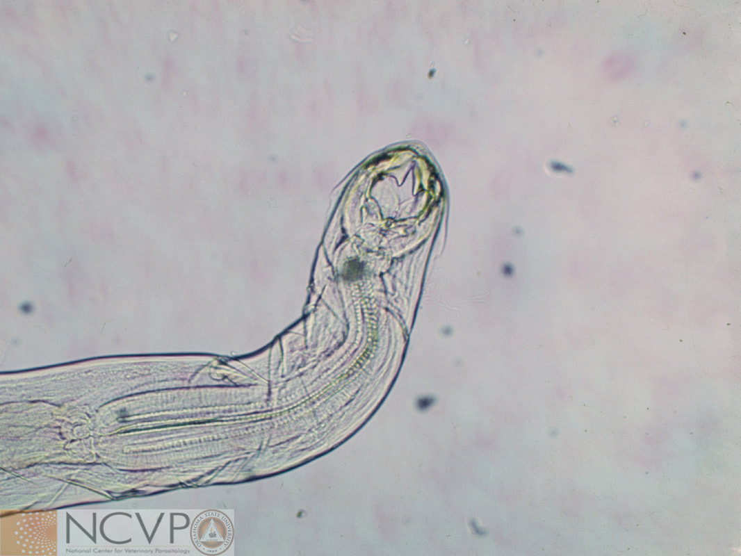
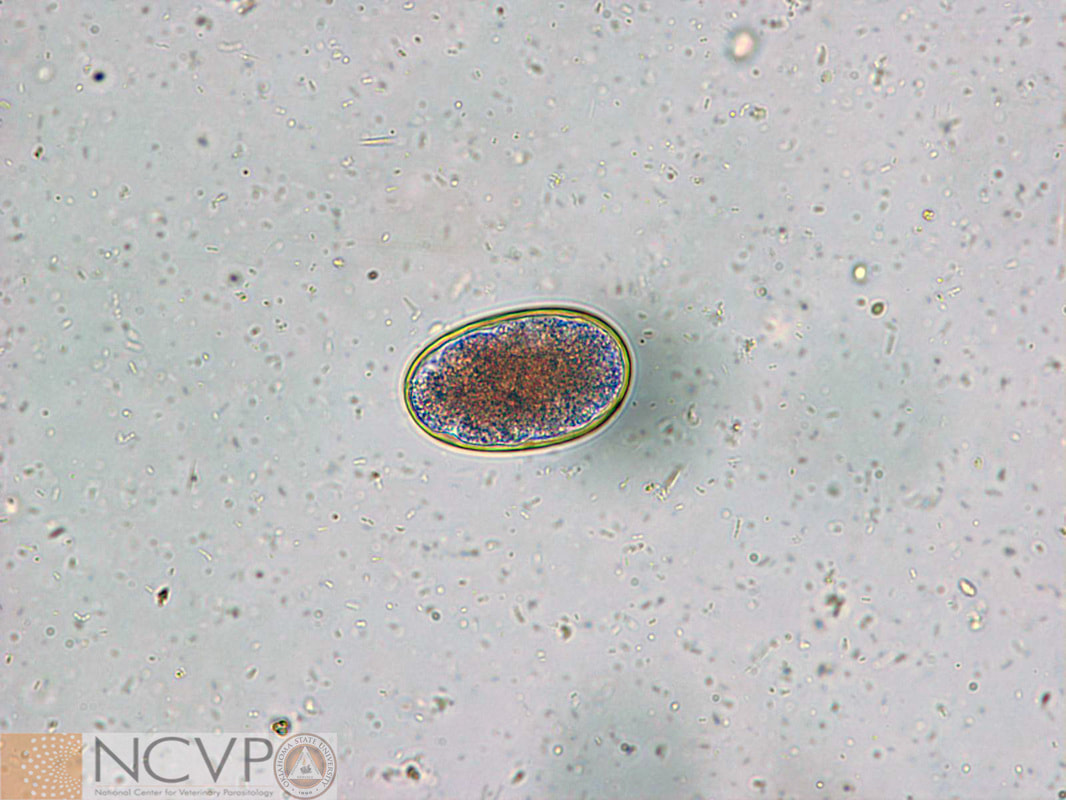
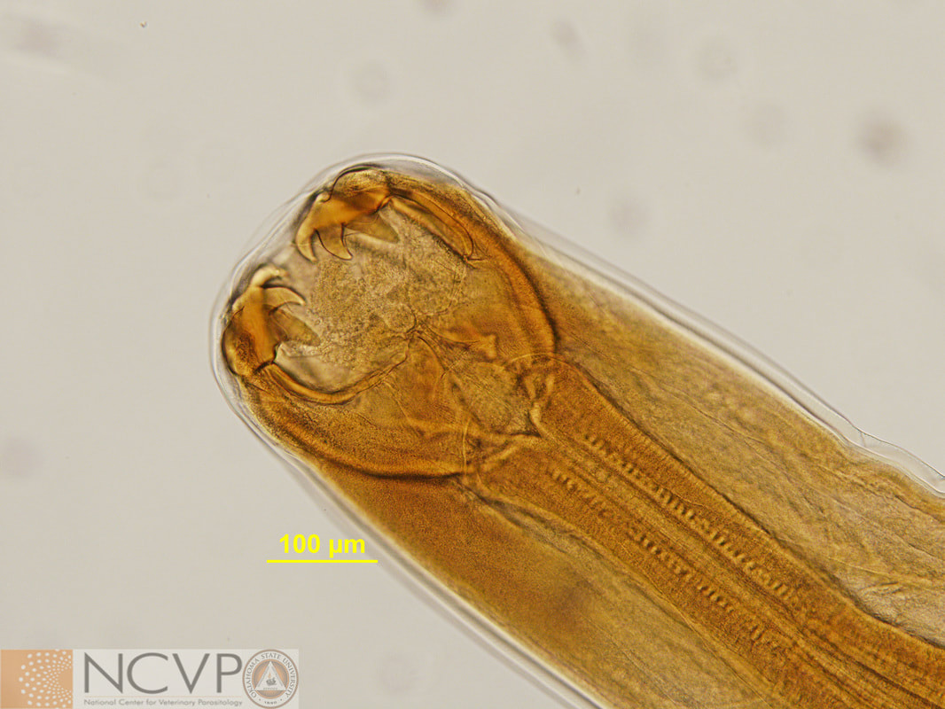
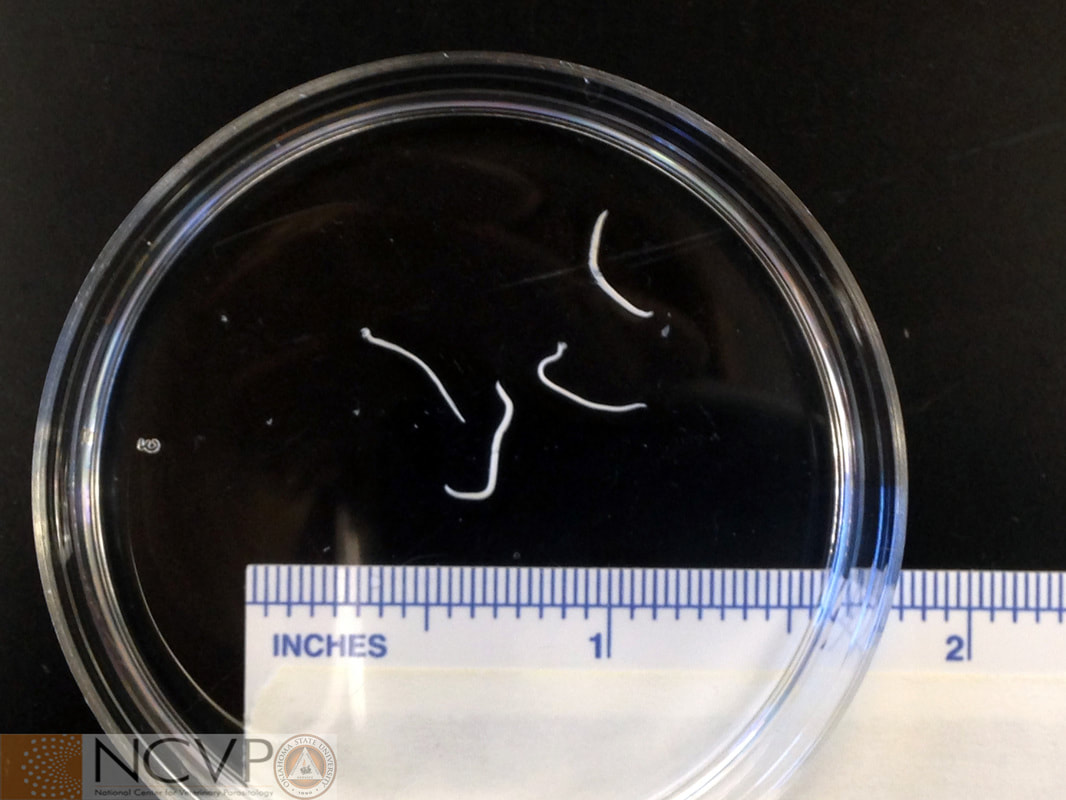
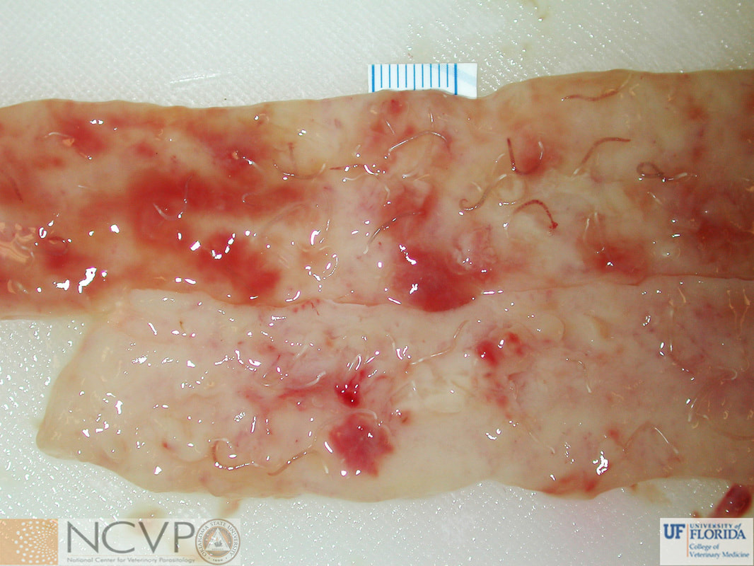
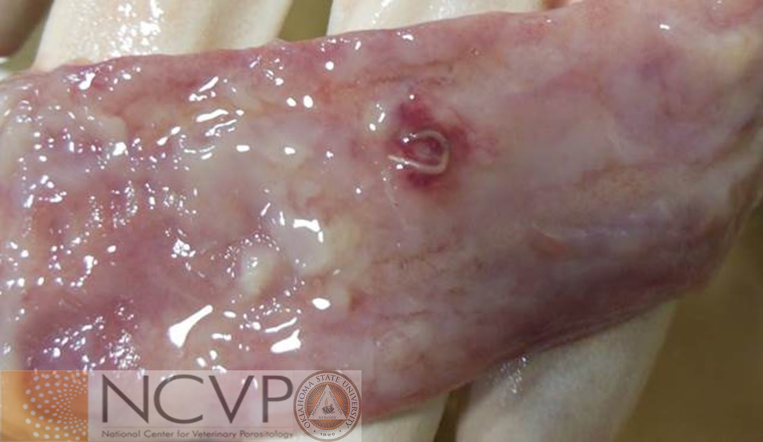
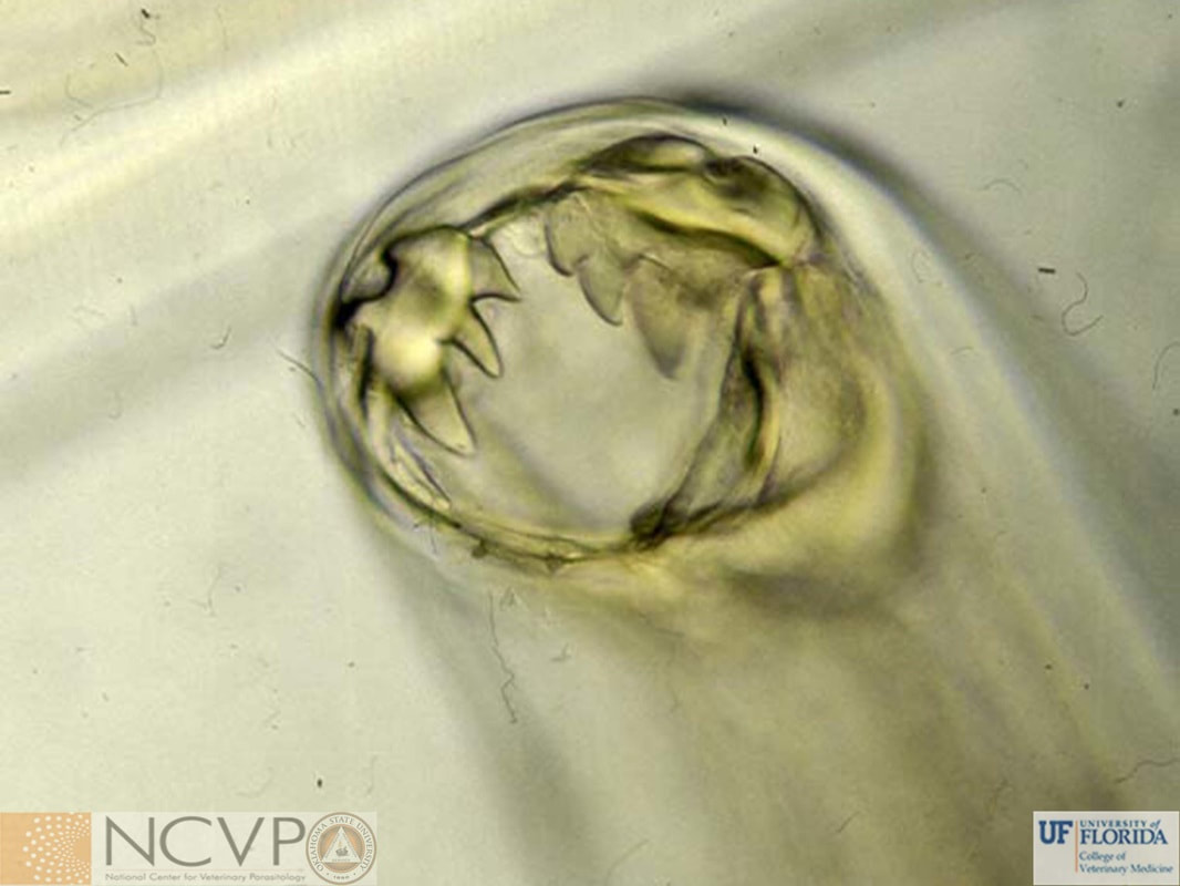
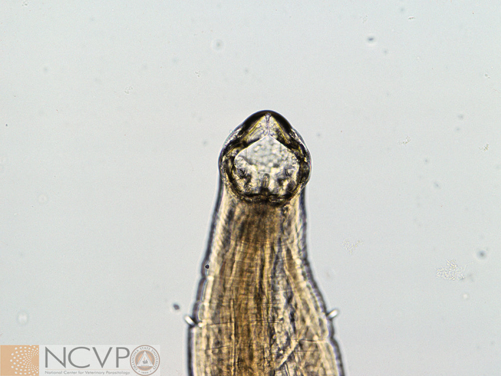
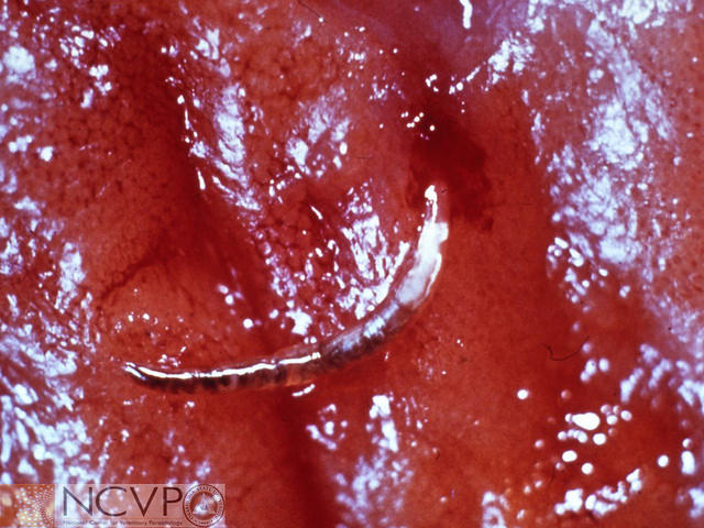
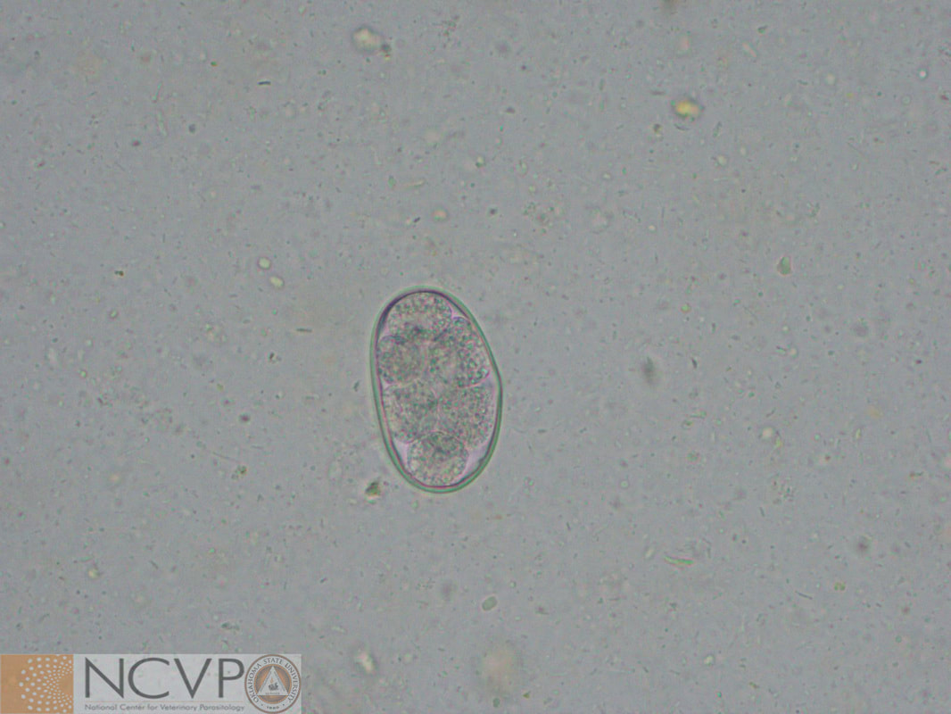
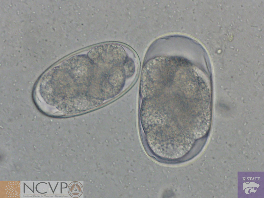
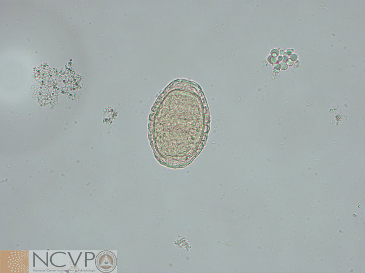
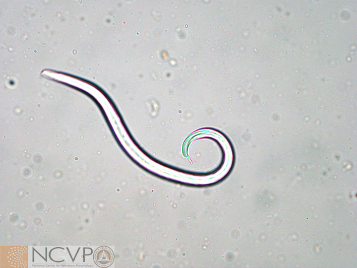
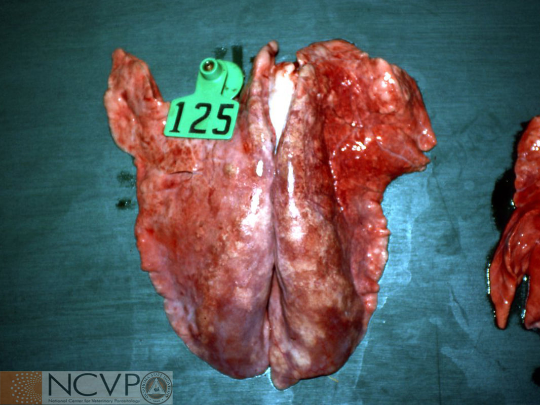
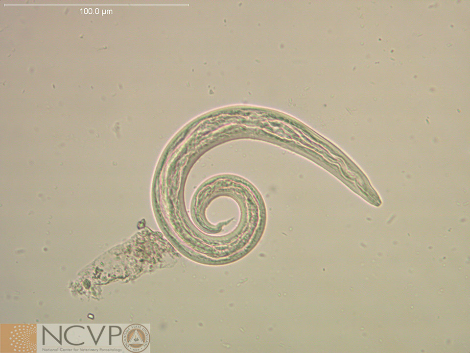
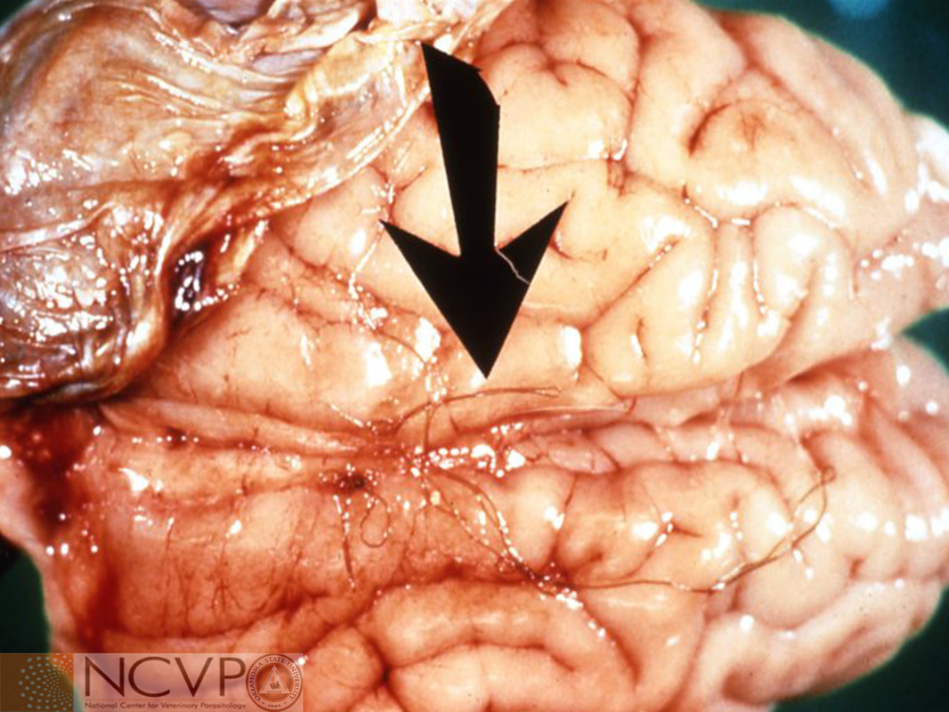
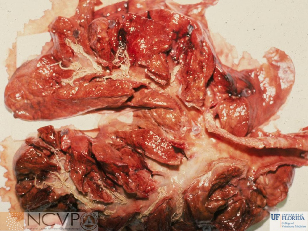
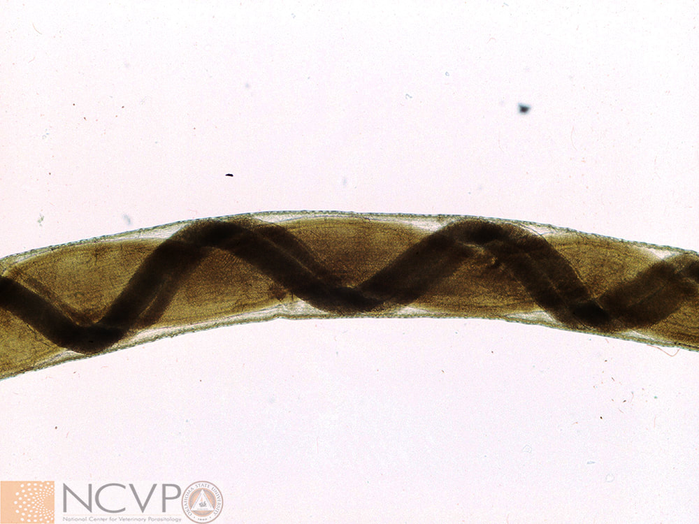
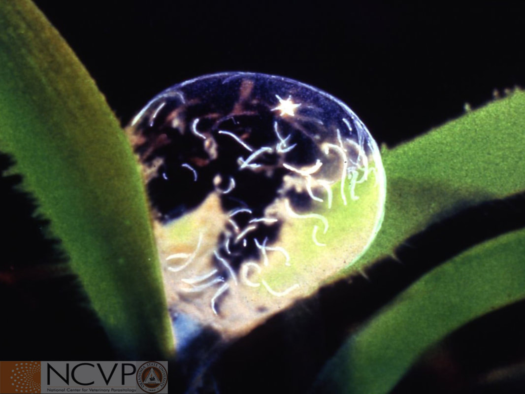
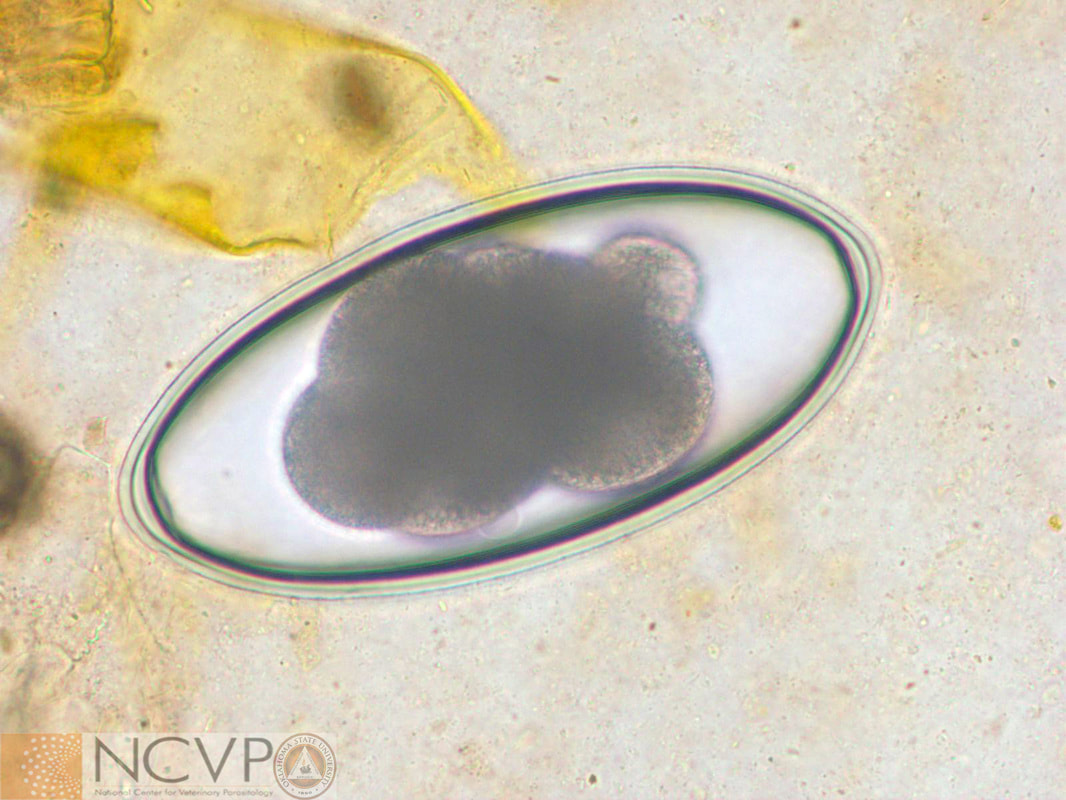
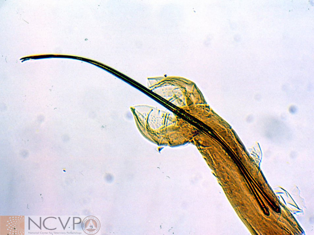
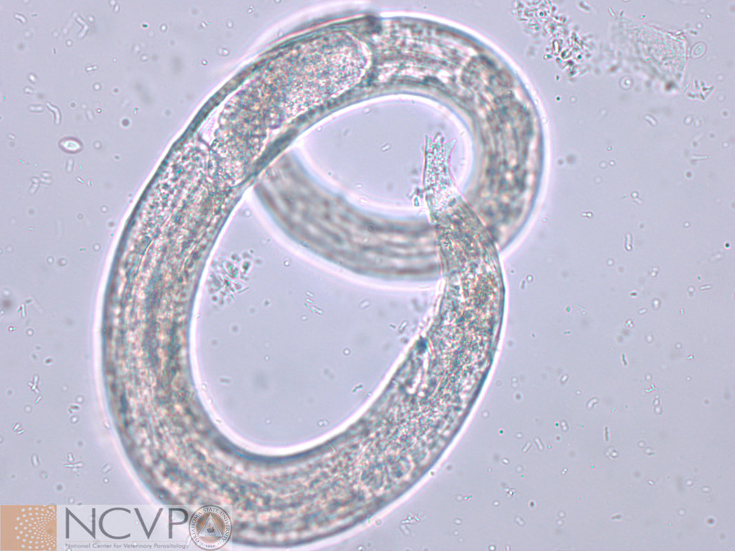
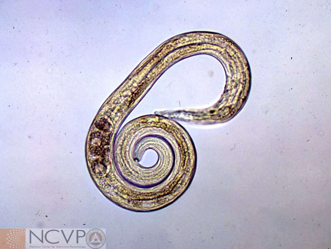
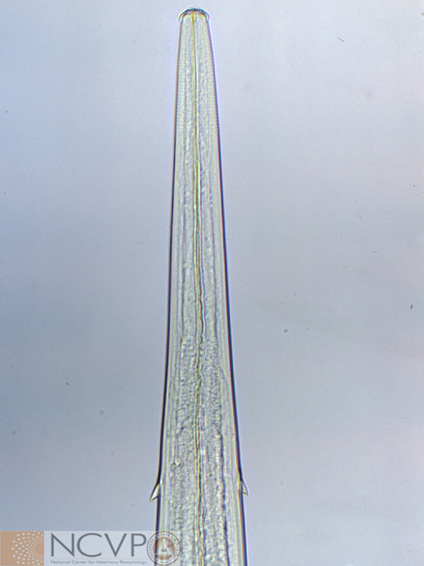
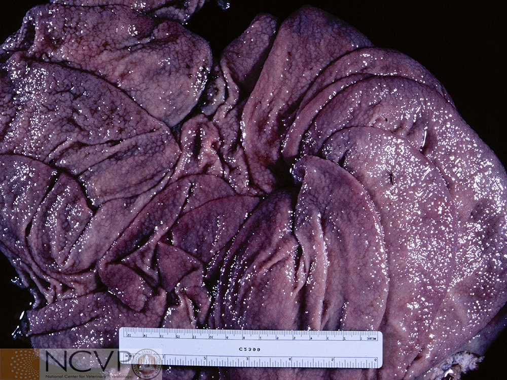
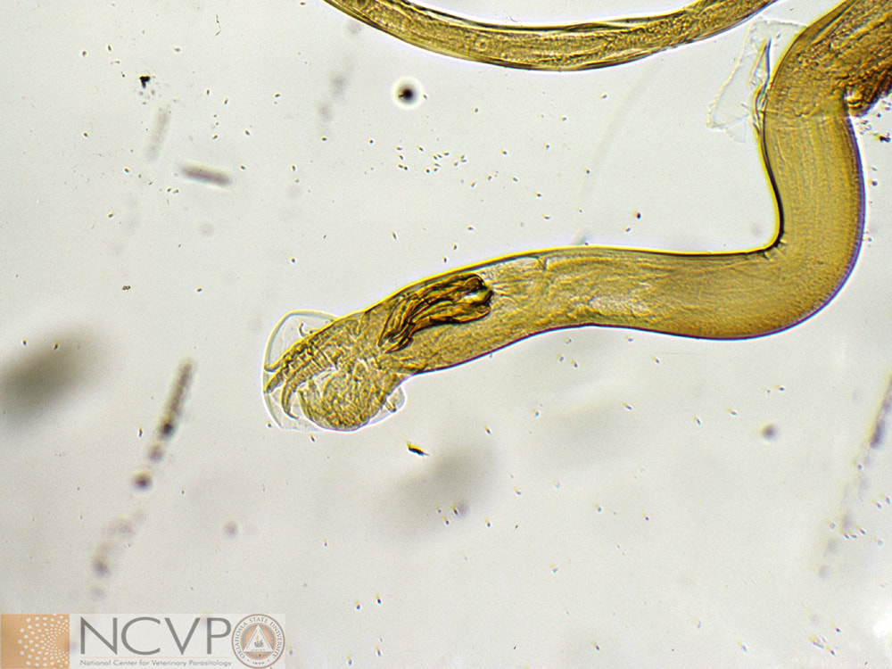
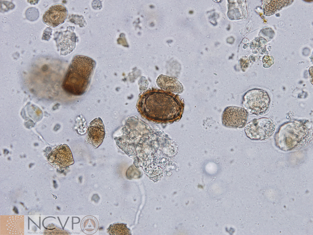
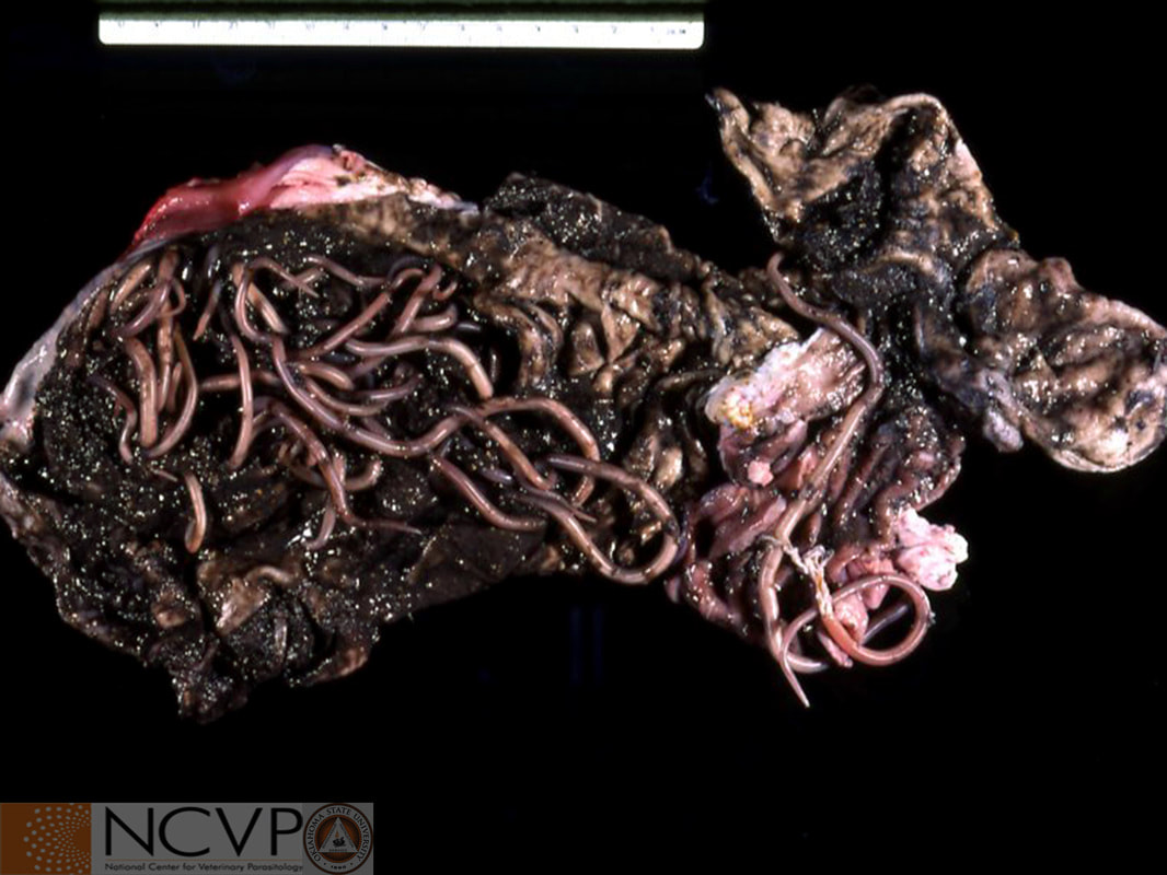
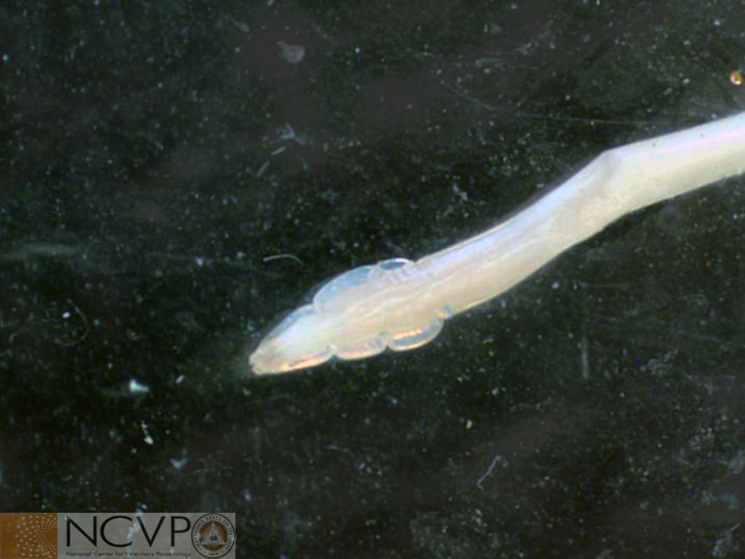
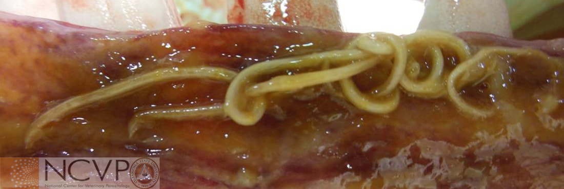
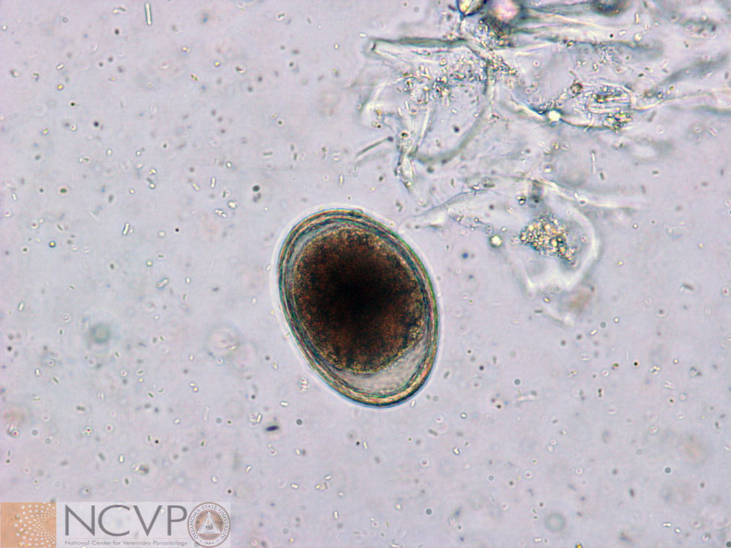
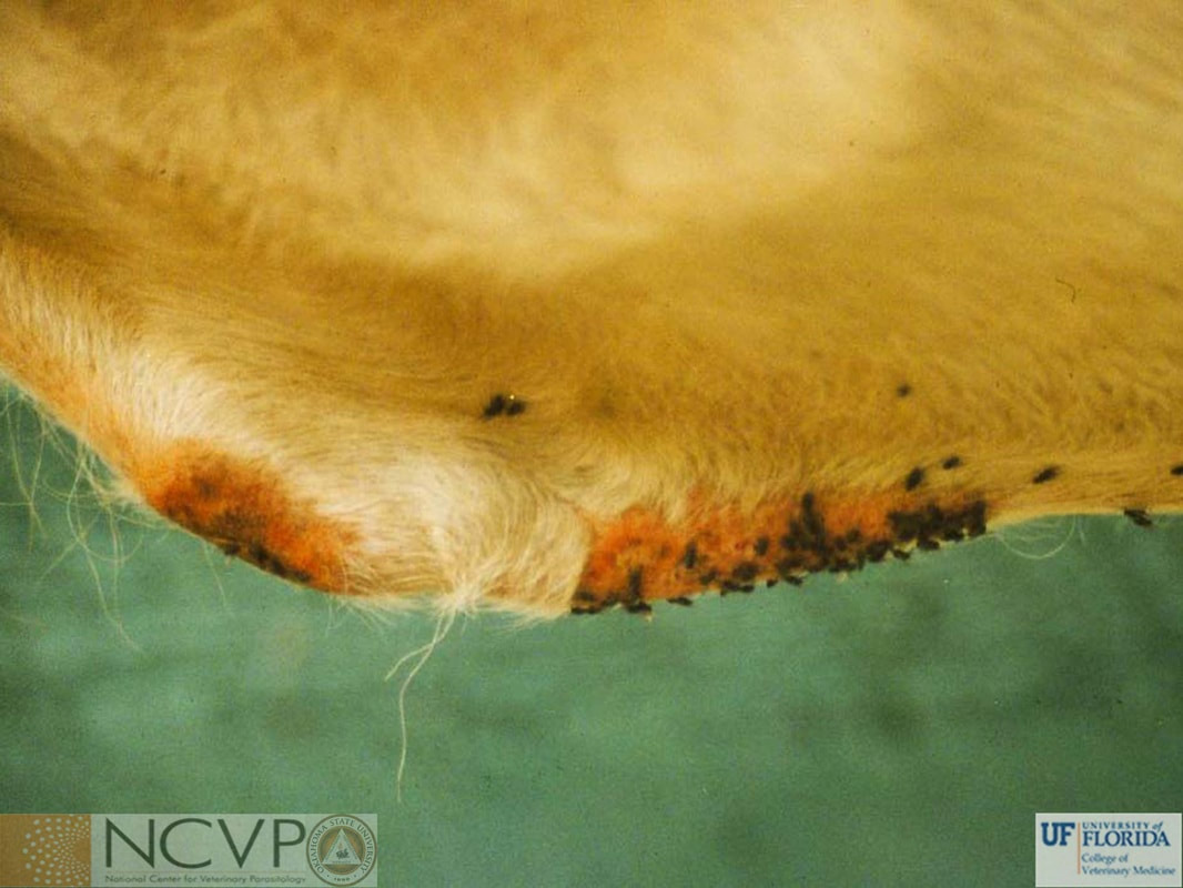
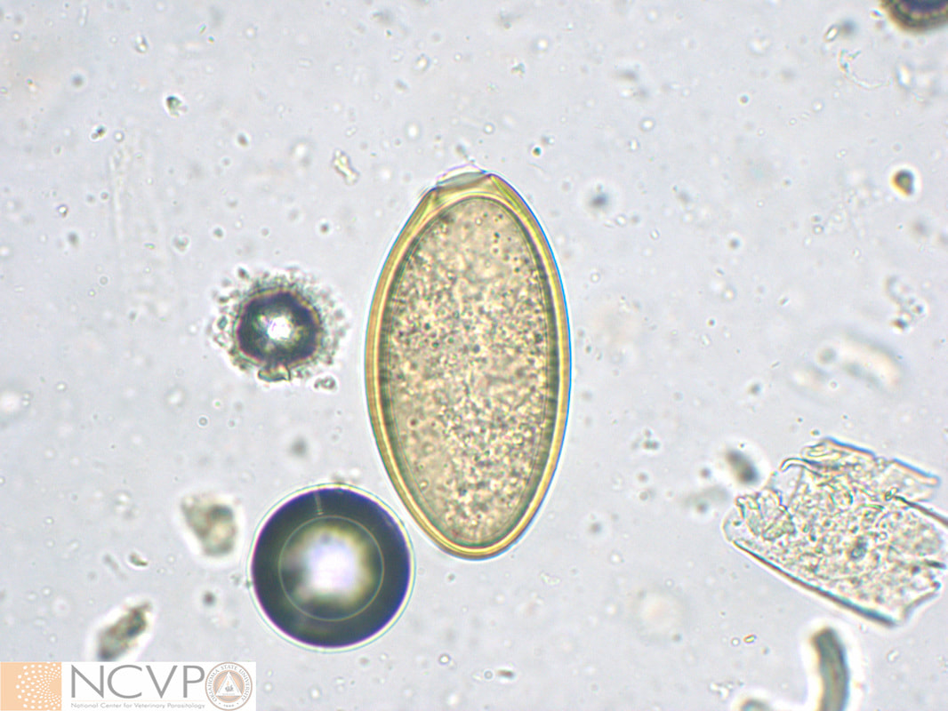
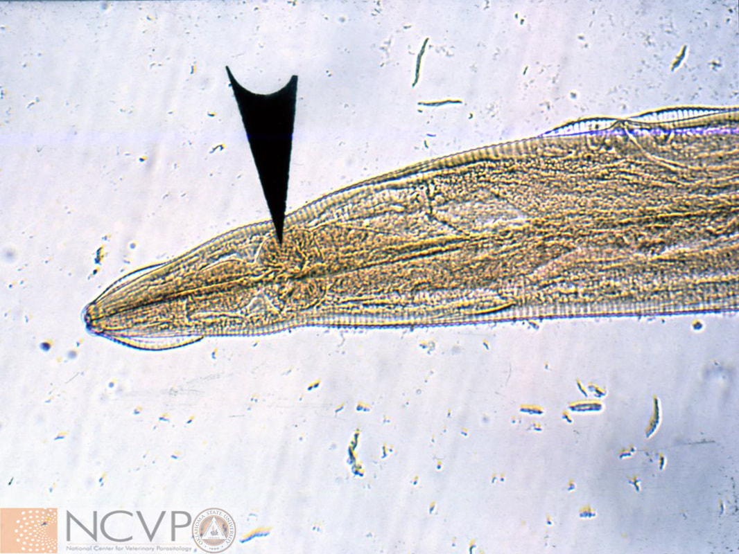
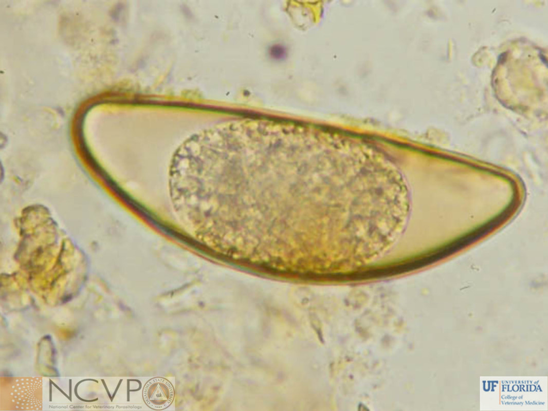
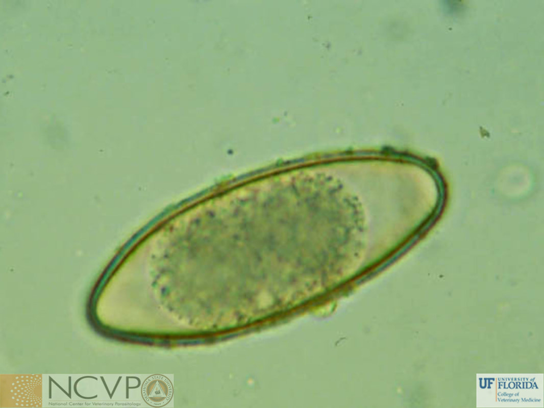
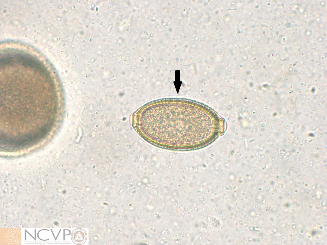
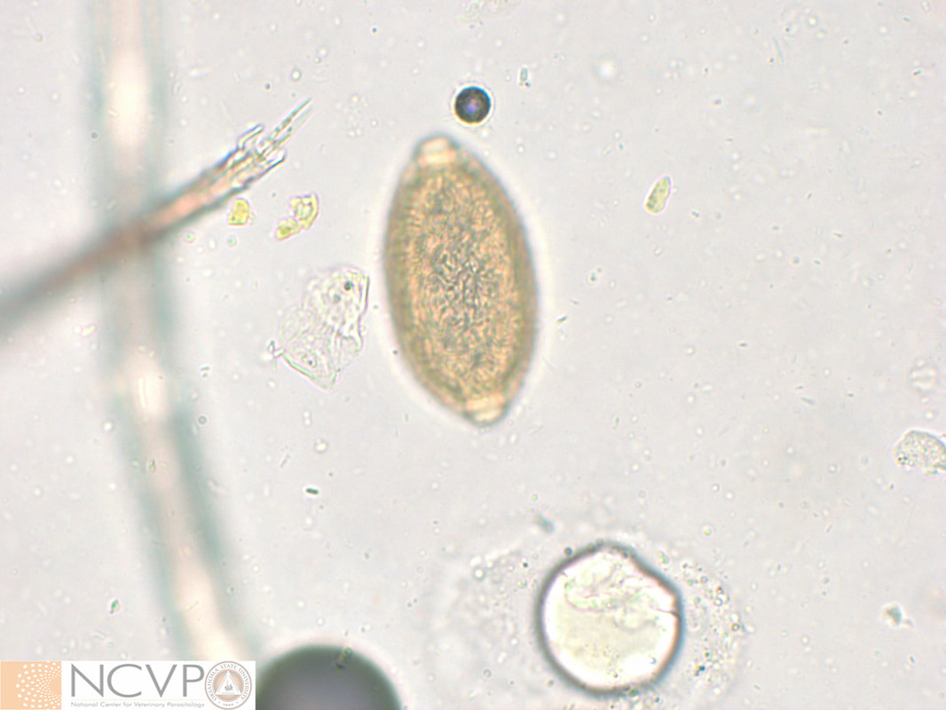
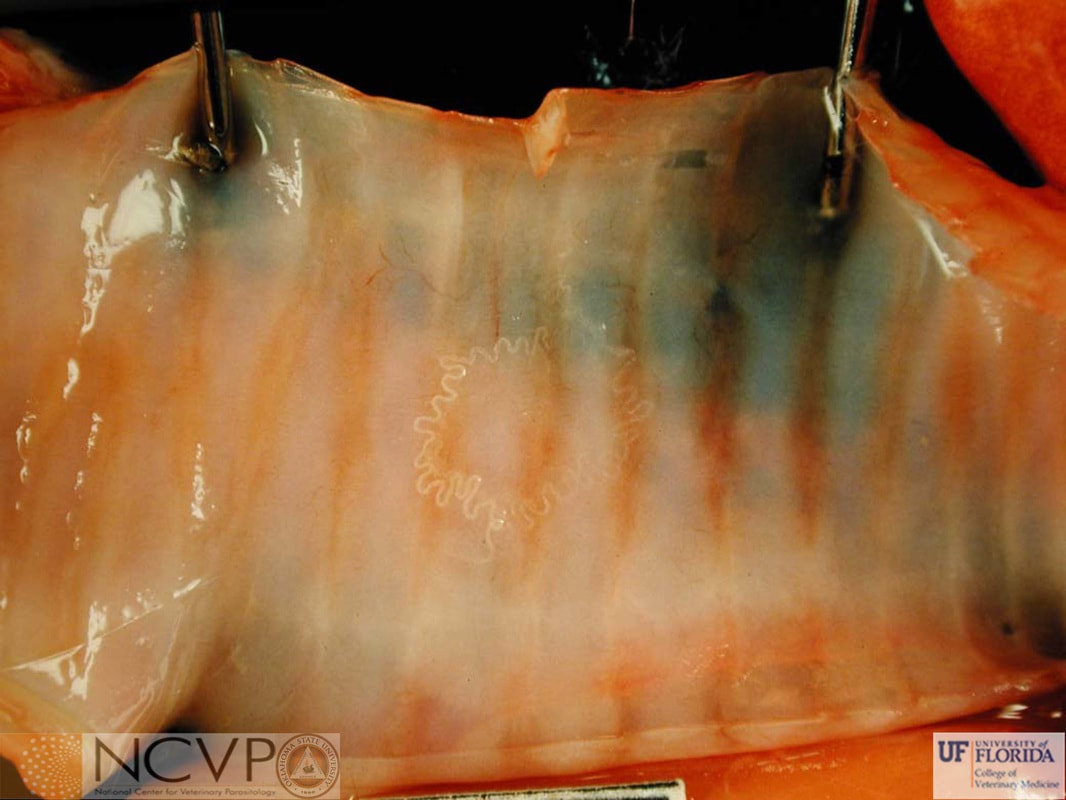
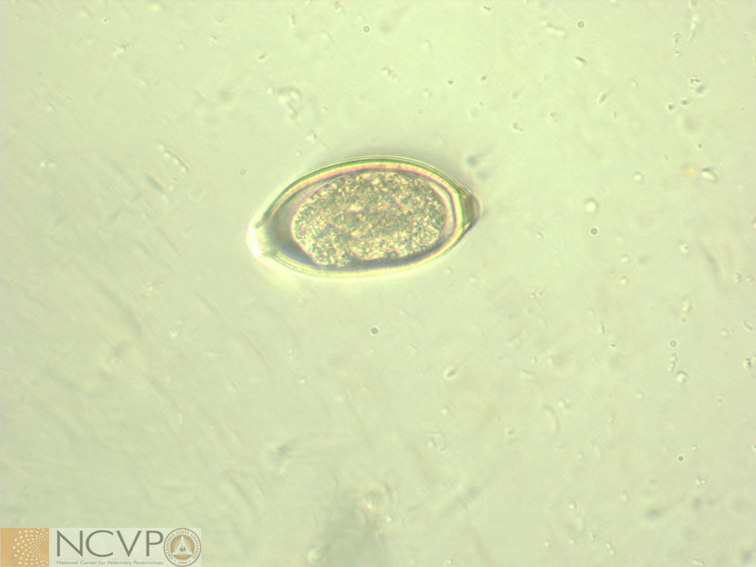
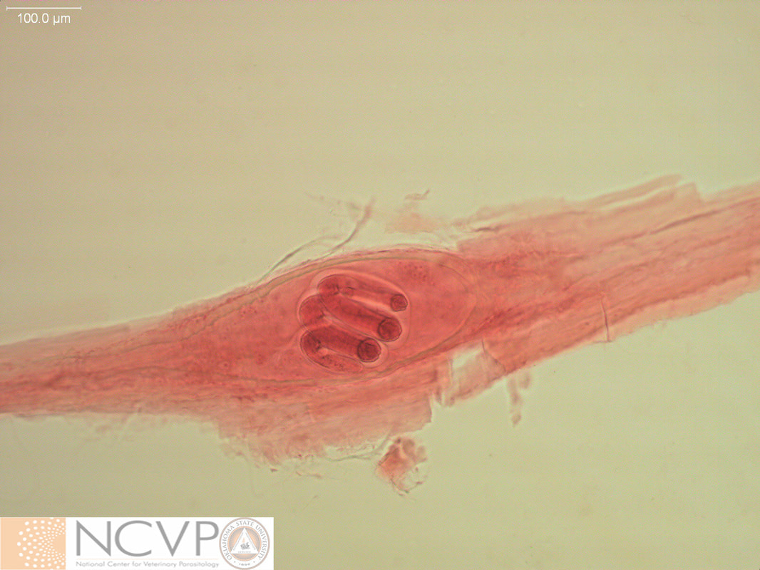
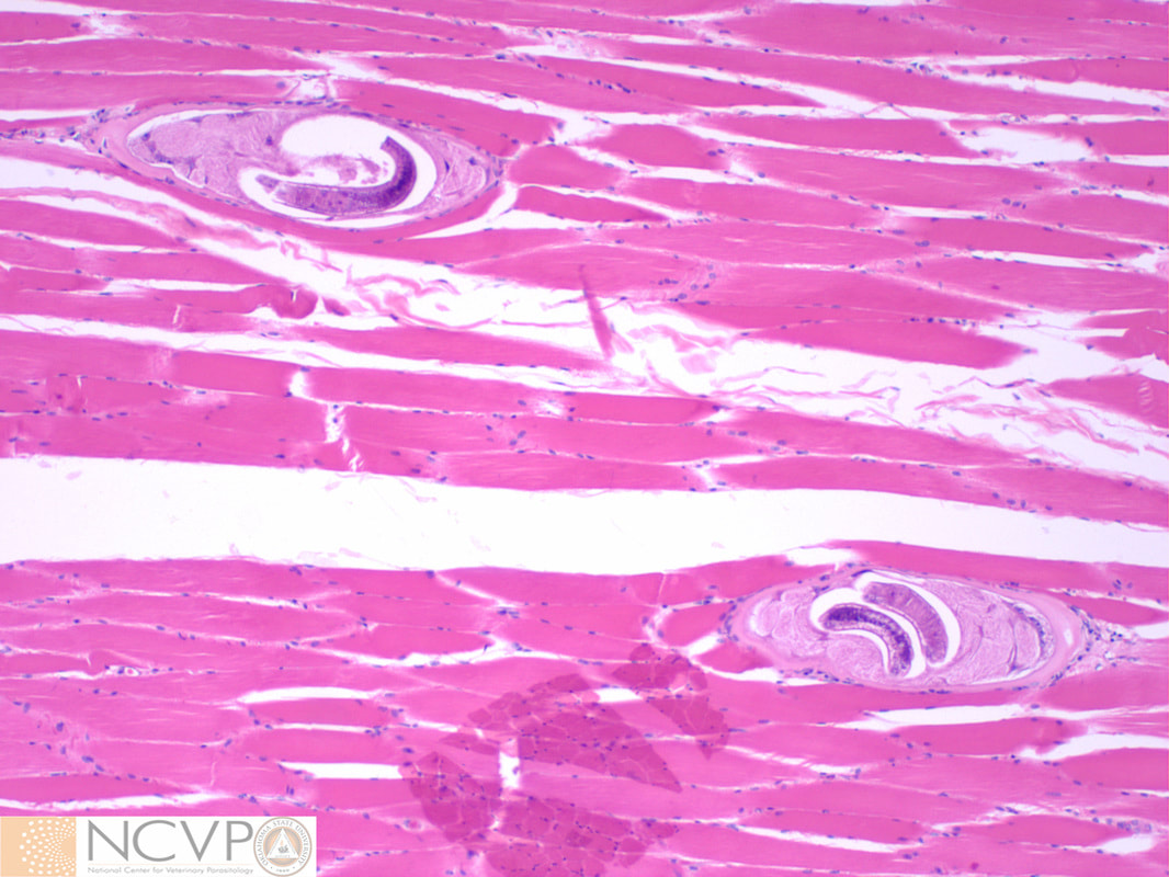
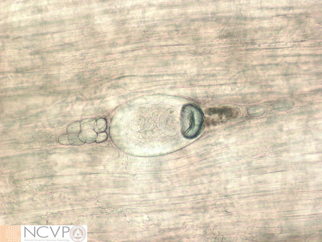
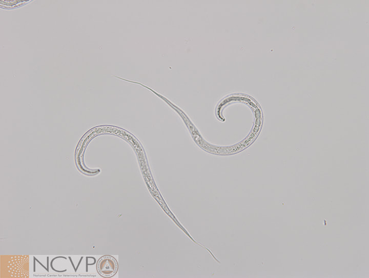
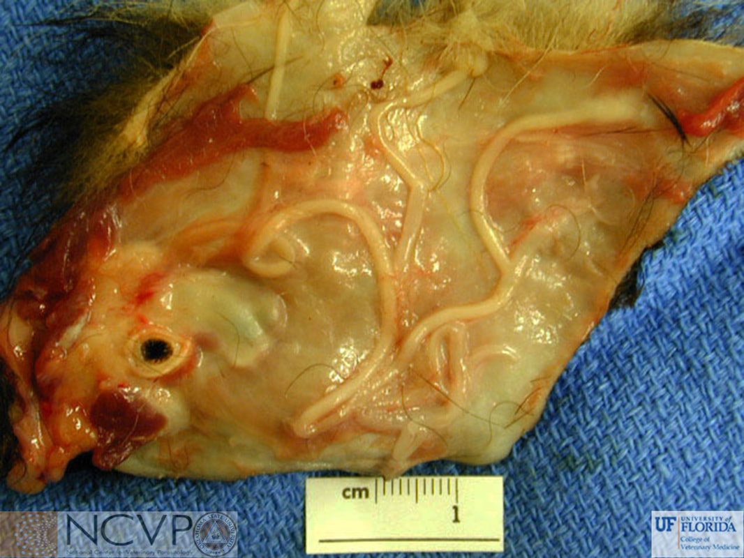
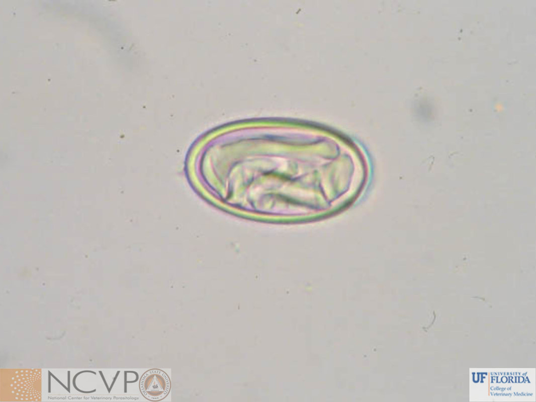
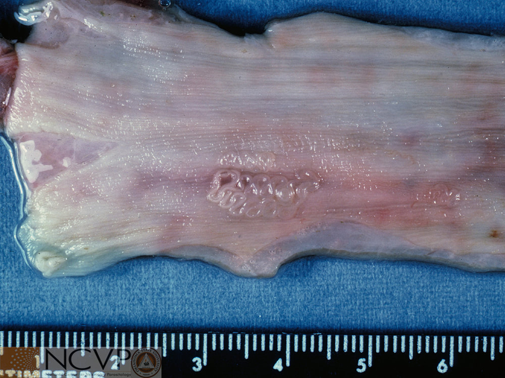
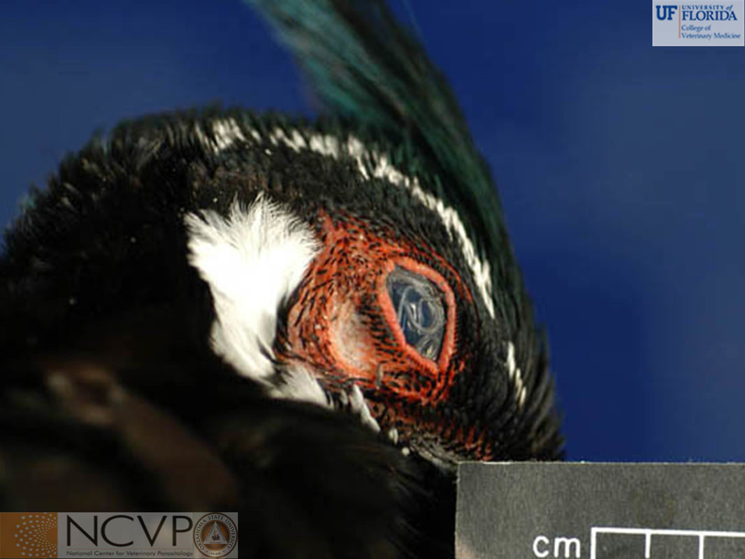
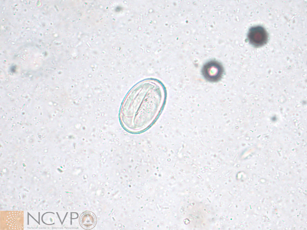
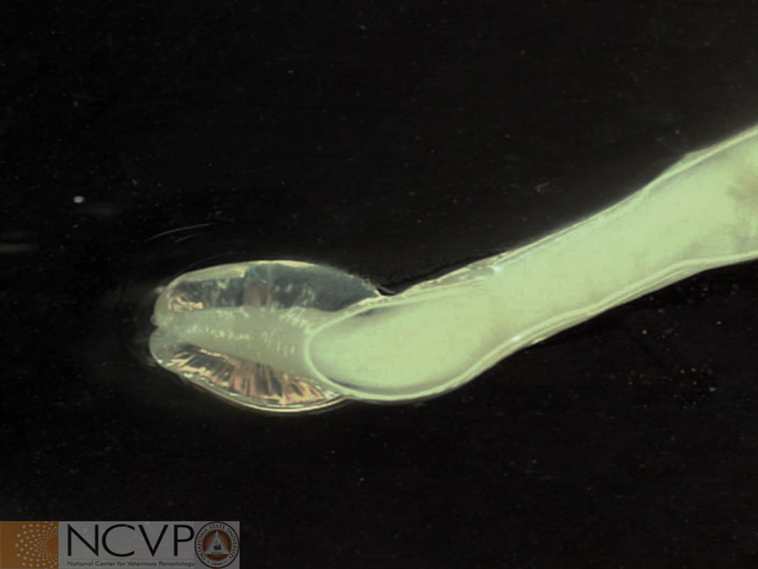
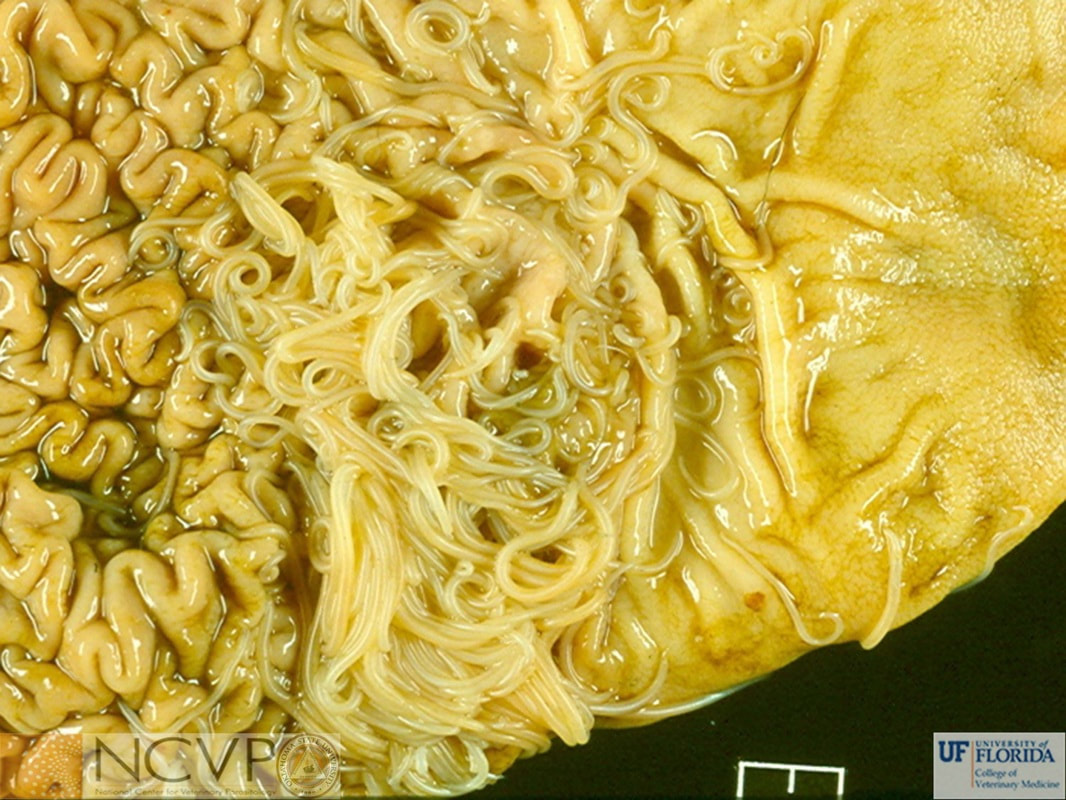
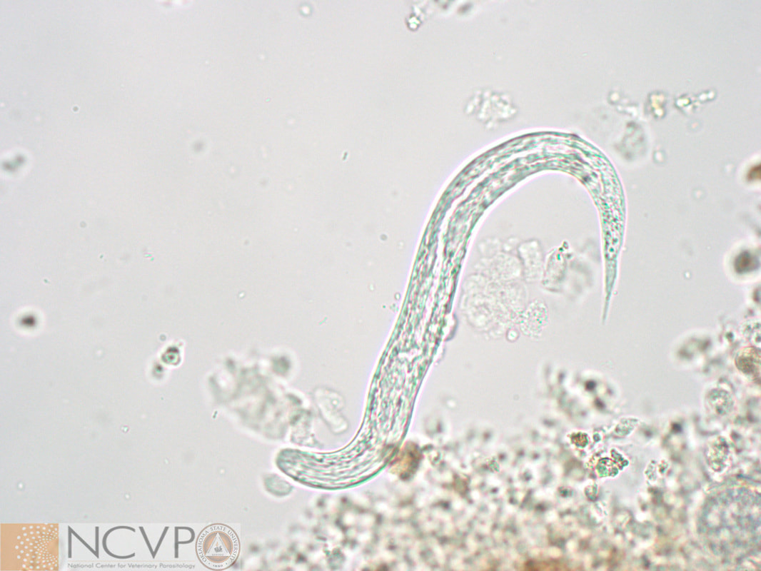
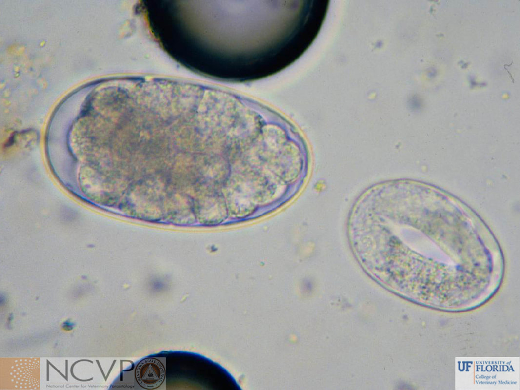
NCVP - National Center for Veterinary Parasitology

  • Home
  • Resources
    • People >
      • Directors and Advisory Board
      • Residents >
        • Boehringer Ingelheim Resident
        • Elanco Resident
        • IDEXX Resident
        • Merck Resident
        • Zoetis Resident
      • NCVP Alums
      • Clinical Parasitology Support
      • Additional Support
      • Sponsors
    • Parasite Image Database >
      • Protozoa
      • Arthropods
      • Nematodes
      • Trematodes
      • Cestodes
      • Acanthocephala
      • Tick-Borne Disease Agents
    • Case of the Month
    • Teaching and Research Materials >
      • Teaching Specimen Request
      • Parasite Jeopardy Games
      • Parasite Videos
    • Online Resources >
      • Parasitology Board Preparation
      • Additional Parasitology Resources
      • Resources for Educators
    • NCVP Board Member Resources
    • NCVP Zoom Backgrounds
    • Residents' Corner
  • News
    • Veterinary Parasitology in the News
    • NCVP Newsletter
  • Opportunities
    • Request for Proposals
    • Residency Application
    • Continuing Education in Parasitology
    • Positions Open in Veterinary Parasitology

Nematodes

Bursate


Ancylostomatoidea - Hookworms

Ancylostoma braziliense
Buccal cavity of adult Ancylostoma braziliense. Note the single pair of cutting teeth.
Ancylostoma caninum
Egg of Ancylostoma caninum in a fecal flotation from a dog. Eggs have an elliptical shape, thin shell, and are approximately 52-79 X 28-58 µm. In fresh feces eggs contain morulae, which develop to first stage larvae within eggs in the environment.
Ancylostoma caninum
Buccal cavity of adult Ancylostoma caninum. Note the three pairs of cutting teeth. Pathogenesis of infection with Ancylostoma caninum is associated with feeding habits of the adult parasite, which leads to blood loss of an estimated 0.01 to 0.09 ml/worm/day.
Ancylostoma caninum
Male and female Ancylostoma caninum recovered from a dog. Adults males are usually 10-12 mm in length, and the females average 14-16 mm in length. Prepatent period varies by route of transmission and can be as short as 2 weeks when larvae are ingested or as long as 4 weeks when larvae are acquired by dermal penetration.
Ancylostoma caninum ​
​In situ photo of Ancylostoma caninum found on necropsy of a dog. Infected dogs may present with anemia from blood loss due to worm feeding, skin lesions associated with penetration of third-stage larvae, diarrhea associated with fourth-stage larvae damaging the small intestine, and/or hemorrhagic pneumonitis associated with larval migration.
Ancylostoma caninum
Adult of Ancylostoma caninum in small intestine from dog at necropsy. Note the area of hemorrhage surrounding the adult.
Ancylostoma tubaeforme
The buccal cavity of the hookworm of cats. Although very similar to the stoma of adult Ancylostoma caninum, the teeth on the ventral margin of Ancylostoma tubaeforme are slightly larger. Infection with this species is the result of skin penetration or ingestion of third-stage larvae, or ingestion of paratenic hosts with larvae encysted in tissues.
Bunostomum sp.
Species of Bunostomum most commonly infect ruminants. A large buccal cavity with cutting plates helps differentiate these hookworms from other intestinal parasites.
​Bunostomum phlebotomum
A parasite of cattle and zebu, Bunostomum phlebotomum may be found in the small intestine feeding on blood. Penetration of larvae may cause cattle to stamp their feet and lick their legs, and heavy infections with adults may lead to diarrhea, anemia, and hypoproteinemia. In calves this manifests as marked weakness, emaciation, and submandibular edema.
Uncinaria stenocephala
Egg of Uncinaria stenocephala. This hookworm infects dogs (rarely cats) in cooler temperate regions, including the northern US, Canada, and Europe. Eggs resemble those of Ancylostoma spp. in that they are elliptical, thin-shelled, and contain morulae in fresh feces, but Uncinaria eggs are slightly larger (71-92 X 35-58 µm).
Uncinaria stenocephala and Ancylostoma caninum
Eggs of Uncinaria stenocephala and Ancylostoma caninum together in a 10X field of view. Note the size difference between eggs of the two species. Photomicrograph provided by Dr. Michael Dryden, Kansas State University.
Uncinaria stenocephala and Ancylostoma caninum
Egg of Ancylostoma caninum on the left and Uncinaria stenocephala on the right in 40X field of view. Photomicrograph provided by Dr. Michael Dryden, Kansas State University.
Uncinaria stenocephala
Buccal cavity of Uncinaria stenocephala. Note the two cutting plates.

Dioctophymoidea

Dioctophyme renale
Eggs of the giant kidney worm, Dioctophyme renale, are passed in the urine of infected hosts. Eggs have a thick, rough shell and measure 60–80 µm by 39–46 µm.
Dioctophyme renale
Adult Dioctophyme renale in the kidney of a dog. Primarily a parasite of mink, other animals, including domestic dogs, other canids, and occasionally pigs, horses, cattle and humans may be infected.
Dioctophyme renale
Adult Dioctophyme renale removed from a coyote. This parasite can also infect humans. Adult female worms may reach 103 cm in length, making Dioctophyme renale the largest nematode known to parasitize humans.

Metastrongyloidea

Aelurostrongylus abstrusus
First-stage larva of Aelurostrongylus abstrusus can be recovered from the feces of infected cats using a Baermann apparatus or zinc sulfate flotation. Larvae are approximately 300–400 µm by 15–20 µm with a terminal S-shaped kink and dorsal spine.
Crenosoma vulpis
Anterior end of Crenosoma vulpis showing the overlapping circular folds unique to the family Crenosomatidae. Adults of this genus are found in the bronchi and trachea of animals. Cresonema vulpis is a cause of substantial chronic tracheobronchitis in captive fox.
Filaroides osleri
Nodules of Filaroides osleri in dog lung. First-stage larvae of Filaroides spp. may be recovered from feces or transtracheal washes. Larvae recovered from feces are 232–266 µm in length whereas larvae from transtracheal washes measure 325–378 µm.
Metastrongylus sp.
Metastrongylus sp. in the bronchus of a pig. Female Metastronglus spp. produce eggs that when passed in feces are larvated and can be detected with flotation.
Metastrongylus apri
Ventral view of the bursa of a male Metastrongylus apri. Adult males may reach 25 mm in length and females may reach 58 mm in length.
Muellerius sp.
First-stage larva of Muellerius sp. recovered from sheep feces. Larvae have an S-shaped kink on posterior end, cuticular spine, and measure 300–320 µm in length.
Muellerius capillaris
Goat lungs showing classic Muellerius capillaris lesions. Adult worms of this genera live in the alveoli and pulmonary parenchyma of sheep and goats. There, they form nodules that turn a greyish color, a result of the degeneration of accumulated leucocytes and necrotic pulmonary tissue. Despite this, hosts generally show no apparent clinical signs.
Parelaphostrongylus tenuis
First-stage larva of Parelaphostrongylus tenuis from a white-tailed deer. Larvae are readily recovered using a Baermann apparatus and measure 310–380 µm in length. Larvae have an S-shaped kink in the tail and a dorsal spine.
Parelaphostrongylus tenuis
Adult Parelaphostrongylus tenuis adults in the subdural space of a white-tailed deer. Adults are 39-91 mm in length.
Parelaphostrongylus tenuis
Neurologic signs of a goat infected with Parelaphostrongylus tenuis. When Parelaphostrongylus tenuis infects a host other than the white-tailed deer, the larvae migrate aberrantly, usually causing damage to the dorsal horns of the grey matter of the spinal cord. Small ruminants are particularly susceptible to neurologic disease due to aberrant migration of Parelaphostrongylus tenuis.
Protostrongylus rufescens
Nodules of Protostrongylus rufescens in sheep lung. Adult Protostrongylus rufescens live in the small bronchioles and may induce nodule formation.

Stephanurus dentatus

Adult Stephanurus dentatus removed from the kidney of a pig. Known as the kidney worm of swine, adults are found in the perirenal fat, the renal pelvis, and the walls of the ureters. Adult males are 20-30 mm and females are 30-45 mm in length.
Adult Stephanurus dentatus excised from cysts in the perirenal fat surrounding the ureter of a pig. Adults are usually found in cysts that communicate with the ureter through which they pass their eggs. The eggs leave the host via urine. Host infections may be acquired through ingestion of third-stage larvae, ingestion of a paratenic host, or from dermal penetration by the third-stage larvae. The larvae then migrate extensively through the organs.

Strongyloidea - Large and Small Strongyles

Strongyle-type egg
Eggs of Strongyloidea have this typical morphology. They are elliptical, have a thin shell, and are approximately 65-100 X 34-50 µm, depending on the species. In fresh feces eggs contain morulae which develop into first stage larvae within eggs in the environment.
Strongyle-type egg
A larvated strongyle egg is shown. Larvated eggs are often seen in fecal samples that have been stored for several days before processing.
Small strongyles
Several different species of adult small strongyles (cyathostomins) from the large intestine of a horse. Over 50 species of equine small strongyles have been described.
Small strongyle
​Buccal cavity of a small strongyle adult. Buccal cavities of small strongyles are smaller than those of large strongyles. Also, note the outer leaf crown on this specimen.
Strongylus spp.
Adult large strongyles (Strongylus spp.) from the large intestine of a horse. The three main species of Strongylus are Strongylus vulgaris, S. equinus, and S. edentatus .
Strongylus edentatus
Strongylus edentatus is differentiated from other members of the genus by a buccal capsule that it is wider anteriorly than at the middle and, as suggested by the species name, a lack of teeth.
Strongylus equinus
The buccal capsule of Strongylus equinus. Strongylus equinus makes an extensive migration through the abdominal cavity, taking 260 days to molt and mature to a patent adult.
Strongylus vulgaris
Buccal cavity of Strongylus vulgaris. Note the two prominent cutting teeth and outer leaf crown.
Strongylus vulgaris
Colic in horses may result from migration of juvenile Strongylus vulgaris through the cranial mesenteric artery.
Gross lesion due to horse strongyles
Thrombosis in the illiac artery of a horse, a result of migrating juvenile strongyles.

Trichonematidae

Chabertia ovina
Large buccal cavity of an adult Chabertia ovina. This parasite is found in the colon of sheep, goats, and cattle throughout the world and feeds on the granular layer that it digests with esophageal secretions. Blood feeding is incidental and may be a cause for frank blood in the feces of the host.
Oesophagostomum radiatum
The nodular worm of cattle, zebu, and water buffalo takes its common name from the characteristic nodules formed around larvae in the intestine of an infected host.
Oesophagostomum columbianum
​Nodules formed by Oesophagostomum columbianum in the colon of a sheep. Oesophagostomum columbianum is a parasite of the colon of sheep, goats, camels, and wild antelope. Their larvae encyst anywhere from the pylorus to the colon to complete their development and become adults that attach in the colon. Frequently, the larval nodules become abcesses that are filled with caseous material.

Trichostrongyloidea

Trichostrongyle-type egg
With the exception of Nematodirus spp., eggs of Trichostrongyloidea species have this typical morphology. They are elliptical, have a thin shell, and are approximately 65-100 X 35-50 µm, depending on the species. In fresh feces eggs contain morulae which develop to first stage larvae within eggs in the environment.
Cooperia spp.
Transverse striations along the cuticle of a Cooperia sp. adult cleared in lactophenol.
​Dictyocaulus spp.
Dictyocaulus viviparus first-stage larva (arrow) recovered from bovine feces. These larvae are approximately 300-360 µm in length and are best recovered using a Baermann apparatus on fresh feces.
Dictyocaulus viviparus
Adult Dictyocaulus viviparus seen in the bronchi of a steer. Dictyocaulus viviparus parasitizes cattle, deer, reindeer, buffalo, and camels. Adults may reach 4-8 cm in length.
Haemonchus spp.
Species of Haemonchus are referred to as barber-pole worms. Their common name reflects the distinct appearance of the female worms - a white uterus wrapped in a spiral fashion around the blood-filled intestines, which appear red in fresh worms.
Haemonchus contortus
Haemonchus contortus are shown feeding in the abomasum of a sheep. When the adults and fourth stage larvae move and leave their initial feeding sites, the wounds continue to bleed into the lumen of the abomasum.
Haemonchus contortus
Third-stage larvae of Haemonchus contortus suspended in a dew drop on grass. Hosts acquire infections with this parasite when they directly ingest larvae from pasture.
Haemonchus contortus
Submitted by Erin Edwards, Texas A&M Veterinary Medical Diagnostic Lab (TVMDL).
2nd Place – Nymphal Place 2020 NCVP Photo Contest.
Haemonchus contortus
Infection with Haemonchus may overwhelm a host's ability to replace proteins and blood lost. This results in decreased intravascular oncotic pressure resulting in edema. The gathering of edema in the submandibular area is termed 'bottle jaw'.
Nematodirus spp.
Egg of Nematodirus sp. Eggs are elliptical, approximately 152-260 X 67-120 µm in size depending on the species, and contain two to eight distinctive large cells in fresh feces. ​
Nematodirus sp.
Spicules and bursa of a male Nematodirus sp. Clinical signs of infection are associated with the emergence of larvae from the mucosa. This manifests as inappetence and acute enteritis.
Ollulanus tricuspis
Larva of Ollulanus tricuspis. Third-stage larvae are approximately 500 µm and have a tricuspid tail similar to that of the adult female (2nd and 4th stage larvae also have this type tail). Adults and larvae are found in the stomach of domestic cats and other felids. Diagnosis is based on the identification of larvae or small adults (1 mm) in vomitus using the Baermann test.
Ollulanus tricuspis
A gravid Ollulanus tricuspis female. In this species, the larvae develop in the uterus of the female until they reach the third stage. Infection is spread when the larvae are vomited and another cat ingests them.
Ostertagia sp.
Anterior end of Ostertagia sp. showing the short buccal cavity and cervical papillae used to differentiate this worm from other trichostrongylids.
Ostertagia ostertagi
Abomasum from a calf infected with Ostertagia ostertagi. The mucosal surface of the abomasum is covered with pin-head to pea-sized nodules as a result of invasion of juvenile worms into gastric glands.
​Trichostrongylus spp.
Adult Trichostrongylus species have a distinct excretory notch visible in the esophageal region.
Trichostrongylus spp.
Species of Trichostrongylus are differentiated from other nematodes by the short and pointed spicules of males.

Non-Bursate


Ascaridoidea - Ascarid Roundworms

Ascaris suum
Egg of Ascaris suum in a fecal flotation from an infected pig. Eggs are 50-70 by 40-60 µm, brownish in color, and surrounded by a thick, rough shell. The prepatent period for this species is 6-8 weeks.
Ascaris suum
Adult Ascaris suum seen in the cecum of a pig on necropsy. Adults of this species infect the small intestine and cecum of pigs and may range from 15 cm to 41 cm in length. ​
Ascaris suum
The characteristic multi-focal blotchy pale regions of this liver from a pig are termed, "milk spots". They are a result of a fibrous response to the migration of immature stages of Ascaris suum through the liver.
Baylisascaris procyonis
Egg of Baylisascaris procyonis, commonly known as the raccoon roundworm. Dogs and raccoons acquire the infection through ingestion of a paratenic host. Ingestion of larvated eggs may cause severe central nervous system or ocular disease in dogs, birds, rabbits, rodents, marsupials, and humans
Baylisascaris sp. and Toxocara canis
A comparison between Baylisascaris sp. (left) and Toxocara canis (right) eggs in a dog fecal flotation. Toxocara canis eggs are larger, measuring 85-90 μm x 75 μm while Baylisascaris sp. range in size from 63-75 μm x 53-60 μm.
Heterakis gallinarum
Anterior end of adult Heterakis gallinarum. Note the large esophageal posterior bulb. Heterakis gallinarum is an inhabitant of the ceca of galliform birds. It is commonly referred to as the "cecal worm."
Heterakis gallinarum
Posterior end of a male Heterakis gallinarum. Note the pre-cloacal sucker and spicules. Heterakis gallinarum is a carrier of Histomonas meleagridis, a protozoal parasite that contributes to blackhead disease in turkeys.
Parascaris sp.
Egg of Parascaris sp. in a fecal flotation from a horse. Eggs of Parascaris equorum and Parascaris univalens are 90-100 µm with a thick, rough shell. After ingestion of eggs, it takes approximately 10 weeks for infections to become patent.
Parascaris equorum
Adult female (top) and male (bottom) Parascaris equorum, a nematode of the small intestine of equids. The males are 15-28 cm in length and the females may reach 50 cm in length. This parasite is most pathogenic to foals 3 to 9 months of age, affecting the respiratory and digestive systems. Adults of the related ascarid Parascaris univalens also infecting equids are considered morphologically identical to P. equorum, but are karotypically distinct.
Toxocara canis
Egg of Toxocara canis in a canine fecal flotation. Eggs are 85-90 by 75 µm, subspherical, and have a thick and pitted shell. After ingestion by the host, it takes 3-4 weeks for the larvae to migrate and mature into egg producing adults.
Toxocara canis
Adults of Toxocara canis from the small intestine of an infected dog. Transmission of Toxocara canis to canids may occur prenatally, through direct transmission, through a paratenic host, or through nursing. Humans may serve as aberrant or parentetic hosts, resulting in cutaneous larva migrans (CLM) and occular larva migrans (OLM).
Toxocara canis
Anterior end of Toxocara canis adult. Note the cervical alae, which are not as prominent grossly as those of Toxocara cati, an ascarid of felids.
Toxocara canis
A puppy with a heavy load of Toxocara canis parasitizing its small intestine. Note the distended belly and constipation. Other common clinical signs include emaciation, dull hair coat, vomiting, and harsh lung sounds associated with secondary pneumonia. Death frequently occurs two to three weeks after birth when pups are prenatally infected.
Toxocara canis
Adults of Toxocara canis in the small intestine of a dog at necropsy.
Toxocara cati
Egg of Toxocara cati in a fecal flotation from an infected cat. Eggs are similar to those of Toxocara canis , but are 65 by 75 µm, and tend to be more elliptical.
Toxocara cati
Toxocara cati adults from the small intestine of an infected cat. The arrowhead shape of the cervical alae is grossly visible.
Toxocara cati
Anterior end of Toxocara cati adult. Note the prominent cervical alae. The alae are easily seen without magnification, unlike those of Toxocara canis.
Toxascaris leonina
Egg of Toxascaris leonina, an ascarid infecting dogs, cats, foxes, and related host species. Eggs are approximately 70-80 µm and resemble those of Toxocara spp., but have a smooth shell and the embryo takes up less space within the egg. ​
​Toxocara vitulorum
Egg of Toxocara vitulorum, the ascarid of cattle. Toxocara vitulorum eggs are subglobular with a finely pitted shell, and measure 75-95 μm x 60-75 μm.

Cruziidae

Cruzia americana
Adult male Cruzia americana. This parasite inhabits the cecum and proximal colon of opossums in the United States. In low numbers they are non-pathogenic, but heavy worm burdens may lead to malabsorption and diarrhea. Males measure 11-12 mm in length and females measure 16-18 mm in length.
Cruzia americana
Egg of Cruzia americana measuring 110 μm x 66 μm. These eggs may be found as pseudoparasites in dog feces if the dog ingested an opossum or its feces.

Filaroidea

Acanthocheilonema reconditum
Fixed and stained microfilaria of Acanthocheilonema reconditum from the blood of an infected dog. Microfilariae of Acanthochilonema reconditum differ from those of Dirofilaria immitis based on size (250-288 µm in length by 4.5-5.5 µm in width), head shape (blunt) and tail shape (curved, or hooked).
Dirofilaria immitis
Fixed and stained microfilaria of Dirofilaria immitis from the blood of an infected dog. Microfilariae are 295-325 µm in length, 5-7.5 µm in width, have a tapered head, and a straight tail.
Dirofilaria immitis
Adults of Dirofilaria immitis, the canine heartworm, recovered from the pulmonary artery of a dog at necropsy.
Elaeophora schneideri
A male Elaeophora schneideri exemplifying its tightly coiled tail. An inhabitant of the common carotid and internal maxillary arteries of deer, elk, and sheep, adult worms measure 6-12 cm in length.
Elaeophora schneideri
​Adult Elaeophora schneideri in the common carotid artery of a white-tailed deer. Adults are usually found in breeding pairs in the arteries. The female releases microfilariae which are usually found in capillaries on the forehead and face. An intermediate arthropod host of either the genus Hybomitra or Tabanus will acquire infection from those locations through blood feeding.
Elaeophora schneideri
Sorehead lesion in a sheep caused by the microfilariae of Elaeophora schneideri. In white-tailed deer, little or no clinical evidence of infection is apparent, but in other hosts, the infections are more pathogenic. In sheep, dermatitis with granulomatous inflammation and pruritus, termed 'sorehead' occurs. In elk, the filarial migration is more aberrant leading to blindness and ischemic necrosis of the brain, muzzle, ears, etc.
Onchocerca cervicalis
Microfilariae of Onchocerca cervicalis in horse tissue section. Microfilariae of ​Onchocerca cervicalis are commonly found in the skin over the linea alba, an anatomical location where the intermediate hosts, Culicoides spp., are known to feed.
Onchocerca cervicalis
Swellings on the withers of a horse caused by adult Onchocerca cervicalis encysted in the nuchal ligament. The adults form tunnels and nodules in the connective tissue, but aside from the nodular swellings, no clinical signs are exhibited.
Onchocerca lupi
Microfilariae of Onchocerca lupi recovered via skin snip from a dog that presented with a subconjunctival granuloma in the eye. Microfilariae of Onchocerca lupiare found in the subcutaneous tissues and are ~100µm in length. O. lupi has been associated with ocular lesions in dogs in the southwestern United States and has also been reported in cats.
Photo by: Guilherme Verocai, Texas A&M University
Stephanofilaria stilesi
Lesion caused by Stephanofilaria stilesi on the navel of a brahman cow. Both adults and microfilariae of Stephanofilaria stilesi may be found in the dermis of these dry pruritic lesions. Haematobia irritans (seen on the lesion here) and Stomoxys calcitrans have both been indicated as being competent intermediate hosts for this filarid.

Oxyuroidea - Pinworms

Oxyuris equi
Eggs of Oxyuris equi are asymmetrical, ovoid, have a polar plug, and measure 85–95 by 40–45 μm. Horses are the only host for Oxyuris equi and the "Scotch-tape test" is more effective for recovering eggs than fecal flotation.
Oxyuris equi
Oxyuris equi females recovered from the large intestine of a horse at necropsy. Adult males are 9-12 mm in length and females may reach 150 mm in length.
Oxyuris equi
Anterior end of Oxyuris equi. The esophageal bulb is indicated by the arrow head. Note that the esophagus is narrow at the middle and the bulb is not distinctly demarcated caudally. ​
Syphacia sp.
Egg of Syphacia sp. found in a fecal flotation of a dog. Syphacia is a genus of pinworms found in rodents. They may be found in the feces of dogs as pseudoparasites if the dog ingested an infected definitive host or the feces of an infected host. Eggs measure 100-142 μm x 30-40 μm with a smooth, clear shell wall.
Aspicularis tetraptera
Egg of Aspicularis tetraptera, a pinworm that inhabits the large intestine of mice. Eggs are shed in the feces and can be detected by centrifugal or simple flotation. Eggs measure 70-98 μm x 29-50 μm with a distinctive double shell wall and narrowed poles.

Trichinelloidea - Whipworms, Capillarids, and Trichinella ​spp.

Eucoleus aerophilus
Egg of Eucoleus aerophilus in a fecal flotation from an infected dog. Eggs are asymmetrical and measure 58–79 by 29–40 μm.
​Eucoleus aerophilus
The surface of an Eucoleus aerophilus egg has a network of interconnecting ridges that can be used to differentiate it from other morphologically similar capillarid eggs.
Eucoleus aerophilus and Trichuris vulpis ​
Comparative photo of Eucoleus aerophilus (left) and Trichuris vulpis (right) eggs found in fecal flotation of a dog. Though very similar in appearance, Eucoleus is asymmetrical and smaller in size.
Eucoleus aerophilus
Eucoleus (Capillaria) aerophilus adult found in the trachea of a dog. This parasite can be found in the trachea, bronchi, and bronchioles of dogs, cats and foxes. Infection in dogs and cats are often sub-clinical, but in farmed foxes, infection is an important cause of bronchopneumonia.
Eucoleus boehmi
Egg of Eucoleus boehmi from an infected dog. Eggs are asymmetrical, contain an embryo that does not fill the interior of the egg, and measure 54–60 by 30–35 μm.
Trichinella spiralis
Stained larva of Trichinella spiralis in muscle from an infected pig.
Trichinella murrelli
Stained histological cross-section of coyote muscle infected with Trichinella murrelli. Morphologically, Trichinella murrelli is indistinguishable from Trichinella spiralis. However, Trichinella murrelli most often infects wild animals and Trichinella spiralis infects pigs.
Trichinella nativa/genotype T6
Tissue press preparation of Trichinella nativa/genotype T6 in nurse cell from the tongue of an infected wolverine.
Trichuris suis
Egg of Trichuris suis in a fecal flotation from an infected pig. Eggs are symmetrical, have plugs at both ends, and measure approximately 50–60 by 21–25 μm.
Trichuris suis
Male and female Trichuris suis recovered from a pig. Note the whip-shaped body characteristic of all Trichuris spp., with the posterior end resembling a bull whip "handle", and the thread-like anterior end (stichosome esophagus) resembling the "lash".
Trichuris trichiura
Egg of Trichurs trichiura in a fecal flotation from an infected baboon. Eggs are symmetrical, have plugs at both ends, and measure approximately 50–55 by 20–25 μm.
Trichuris vulpis
Egg of Trichuris vulpis in a fecal flotation from an infected dog. Eggs are symmetrical, have plugs at both ends, and measure approximately 72–90 by 32–40 μm.
​Trichuris vulpis
Adult Trichuris vulpis seen in the cecum of a dog. These whipworms measure 45–75 mm in length and may be found in the cecum and large intestine of dogs and foxes. ​
Trichuris vulpis
Adult Trichuris vulpis in cecum of naturally infected dog.

Spiruroidea

Dispharynx spiralis
Egg of Dispharynx spiralis recovered form the excrement of a pigeon. Dishparynx spiralis is a parasite of birds, occurring in the wall of the proventriculus and esophagus. Eggs are 33-40 μm x 18-25 μm. Infection of the definitive host occurs after ingestion of an infected isopod that serves as an intermediate host.
Dispharynx spiralis
Adult Dispharynx spiralis. The undulating appearance of the anterior end is due to the sinuous, recurrent, course of the cordons that do not anastomose. Males measure 7-8.3 mm in length and females measure 9-10.2 mm in length.
Dyspharynx spiralis
Adult Dyspharynx spiralis in a peacock proventriculus. Pathogenicity is limited to inflammation and thickening of the proventricular lining in light infections, but in severe infections, deep ulcers and extensive destruction of glands lead to weight loss and anemia in young birds.
Dracunculus insignis
Removed from the uterus of female worms, the long, thin tails of first stage Dracunculus insignis larvae are distinctive and measure about 600 μm in length. Intermediate stages are found in copepods (Cyclops) and migrating through the abdomen and thorax of a definitive host (numerous wild carnivore species including raccoons, fisher, mink, muskrat, opossums, skunk, fox, coyote, otter, and occasionally domestic dogs and cats). ​
Dracunculus insignis
Female Dracunculus insignis found in the subcutaneous tissues of a skunk. Dracunculus insignis is a parasite of carnivores in North America. Adult females migrate to the subcutaneous tissues where they form an ulcerative opening in the skin through which they discharge larvae into water.
Gongylonema spp.
Egg of Gongylonema spp. Eggs of this genus are passed in the feces of the infected host. In order to complete its life cycle, it is then swallowed by coprophagous beetles or cockroaches where it develops into the infective stage in about 30 days. It may establish infection when the definitive host ingests an infected insect.
Gongylonema pulchrum
Gongylonema pulchrum infects ruminants, pigs, and less frequently equids. It has also been reported in humans. Adult worms weave sinusoidal tracts within the esophageal mucous membrane.
Gongylonema verrucosum
Adults of Gongylonema verrucosum. This species occurs in the rumen of sheep, goats, cattle, deer, and zebu. Males measure 32-41 mm long and females measure 70-95 mm long.
Habronema sp. / Draschia megastoma
Larvated eggs of Habronema or Drachia megastoma found in horse feces (the species are morphologically indistinguishable). After larvated eggs are passed in the feces, they are ingested by fly larvae and develop into infective larvae. Flies then deposit the third-stage larvae around the lips of horses. The larvae migrate to the horse's mouth and then to the stomach.
Habronema sp. / Draschia megastoma and Gasterophilus sp.
Nodule in horse stomach formed by adult Habronema and/or Draschia megastoma next to larvae of Gasterophilus. Both Habronema and Draschia adults conglomerate and form tumor like nodules in the stomachs of horses where they live and breed.
Habronema sp. / Draschia megastoma
Incised stomach nodule revealing caseous material and adult Habronema/Draschia. Although clinical signs are non-specific, adults may cause gastritis, resulting in poor growth and development of the host.
Habronemiasis
Granulation tissue caused by larvae of Habronema in a non-healing wound on the face of a horse. When flies deposit third-stage larvae of Habronema on open wounds, the larvae prevent the wound from healing. Called, 'summer sores,' these lesions persist during the time that flies are active.
Oxyspirura sp.
Adult Oxyspirura sp. seen under the nictitating membrane of a Palawan peacock. The adults of this genus measure 10-19 mm long. They deposit their eggs into the nasolacrimal duct which are passed into the digestive tract. As infective larvae are liberated from ingested infected cockroaches, they travel back up the bird digestive tract and nasolacrimal duct to reach the eye.
Physaloptera sp.
​Egg of Physaloptera sp. in a canine fecal flotation. Eggs are 42-53 by 29-35 µm, elliptical, have a smooth shell wall, and contain a coiled larva. The eggs mature to the infective stage in an intermediate host (cockroaches, crickets, or beetles).
Physaloptera sp.
Anterior end of Physaloptera sp. adult. Note the cuticular collar and two pseudo lips.
Physaloptera sp.
Posterior end of a Physaloptera male. Note the pronounced caudal alae.
Physaloptera sp.
Adult Physaloptera found in the stomach of a dog. All species of this genera inhabit the proximal half of the digestive tract and adults may be found in vomit. Relocation of feeding sites may cause bleeding and ulceration, resulting in vomiting, anorexia, and melena.
Spirocerca lupi
Eggs of Spirocerca lupi. Though very similar to those of Physaloptera, Spirocerca lupi eggs (30-38 μm x 11-15 μm) are smaller and more elongated than Physaloptera (42-53μm x 29-35μm). These eggs may be found on sedimentation and are found inconsistently on routine fecal flotation exams.
Spirocerca lupi
Adult Spirocerca lupi. Adult Spirocerca lupi are found in nodules in the esophagus, stomach, and aorta of canines and wild felines such as bobcats. The eggs are passed in the feces, where they are ingested by a coprophagous beetle intermediate host in which they will become infective. After the beetle is ingested by a definitive host, the larvae extensively migrate through the arteries and aorta until reaching the esophagus.
Spirocerca lupi
Adult Spirocerca lupi excised from and pictured next to its nodule, found in the stomach of a dog. Complications associated with Spirocerca lupi include dysphagia, occulusion of esophagus, spondylitis, malignant tumor development, aneurysm, and focal necrosis.
Thelazia sp.
A species of Thelazia and associated pathology in the eye of a harvested mule deer. Thelazia species inhabit the conjunctival sac or lacrimal duct of mammals and birds. Musca autumnalis serves as an important vector for Thelazia in the United States.

Rhabditoidea

Strongyloides papillosus
Egg of Strongyloides papillosus found in an ovine fecal preparation. These eggs are elliptical with a thin shell, 40-60 X 32-40 μm, and in fresh feces, contain first-stage rhabditiform larvae. Rhabditiform refers to the shape of the esophagus.
Strongyloides stercoralis
Third stage larva of Strongyloides stercoralis. This is the infective form which may burrow through the skin of the host and travel in the blood to the lungs. From the lungs, it migrates to the trachea where it is coughed up and swallowed, passing through the stomach and establishing its infection in the small intestines of carnivores.
Strongyloides sp.
In fresh feces, Strongyloides spp. larvae rapidly develop to the infective filariform stage, which enters the host via skin or mucosal penetration. Filariform refers to the elongate shape of the esophagus. In dogs and cats, Strongyloides eggs frequently hatch before leaving the body, thus free larvae are most often found in fresh feces.
Strongyloides westeri and strongyle sp. egg
Comparative image of a strongyle egg (left) and Strongyloides westeri larvated egg (right) found in an equine fecal float. ​
Strongyloides westeri
In foals as young as 10-14 days old Strongyloides westeri may cause acute diarrhea. An age related immunity is developed at 15-23 weeks of age for horses but may be delayed to 9-12 months in donkeys.

    Categories

    All
    Bursate
    Non-Bursate