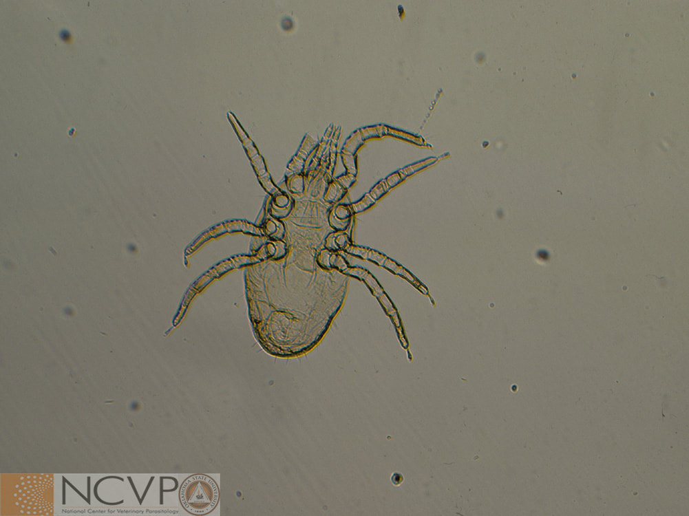
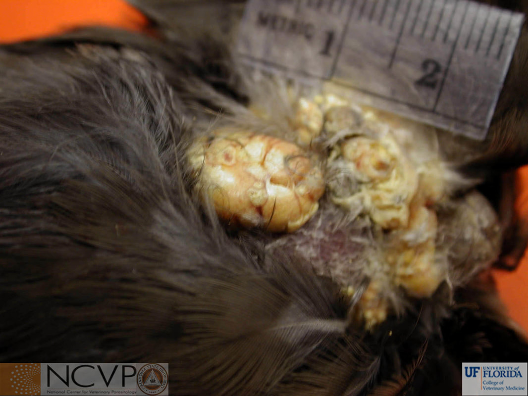
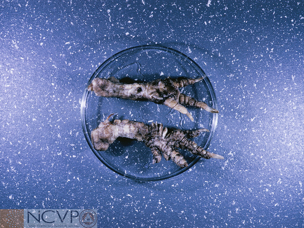
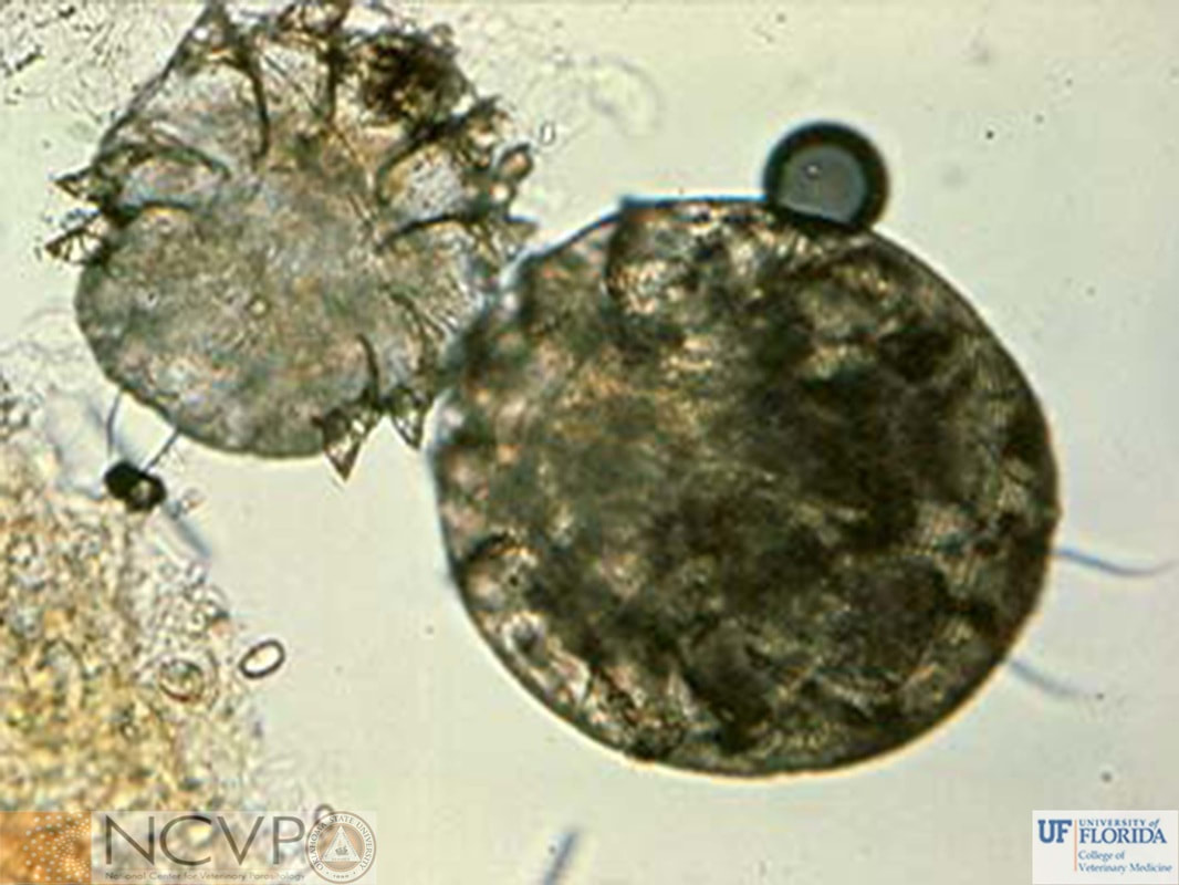
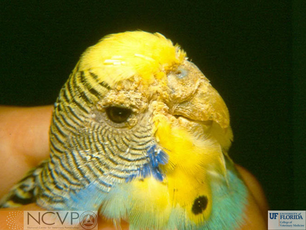
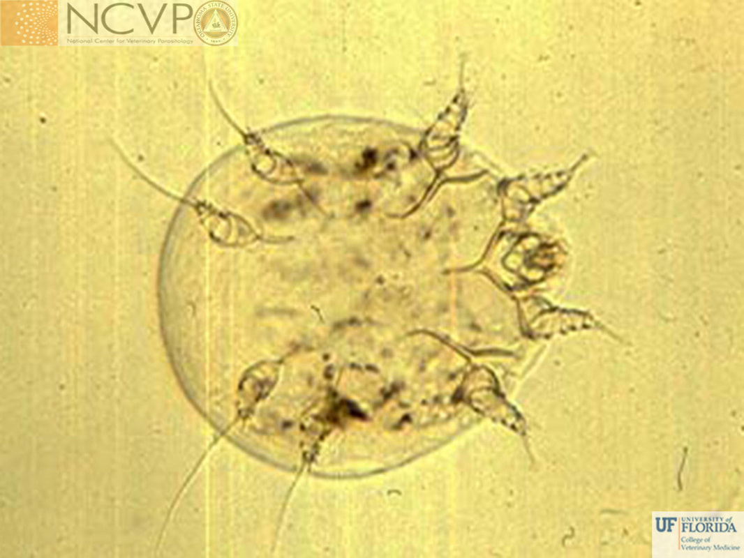
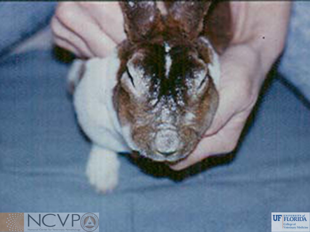
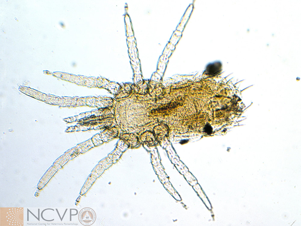
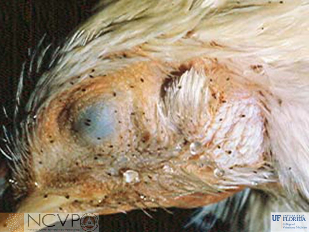
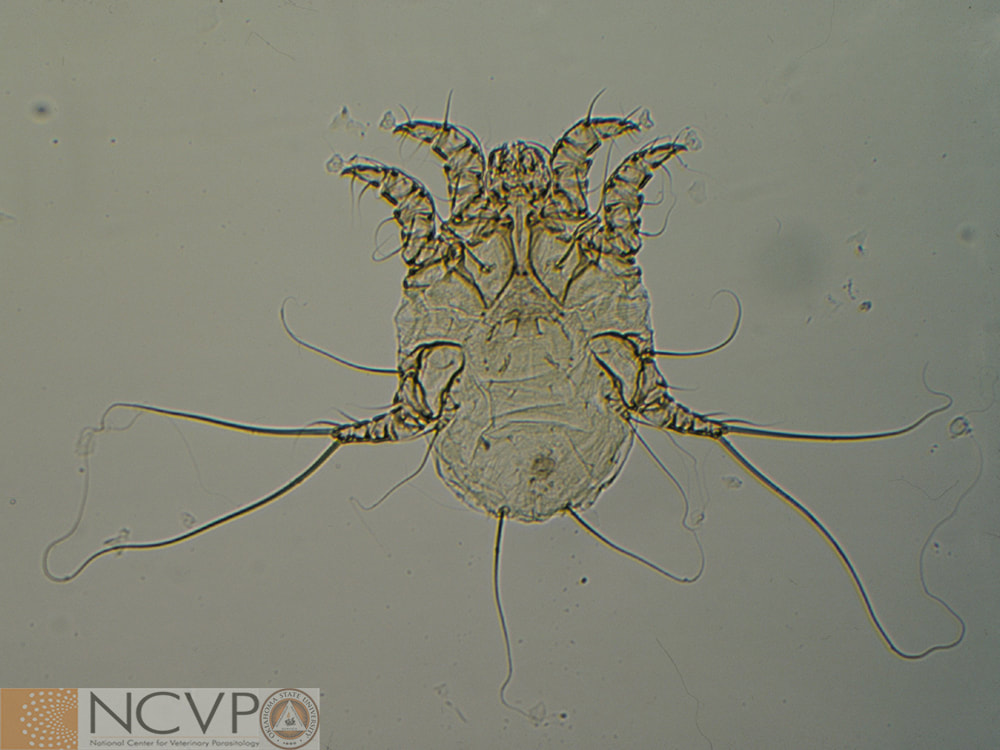
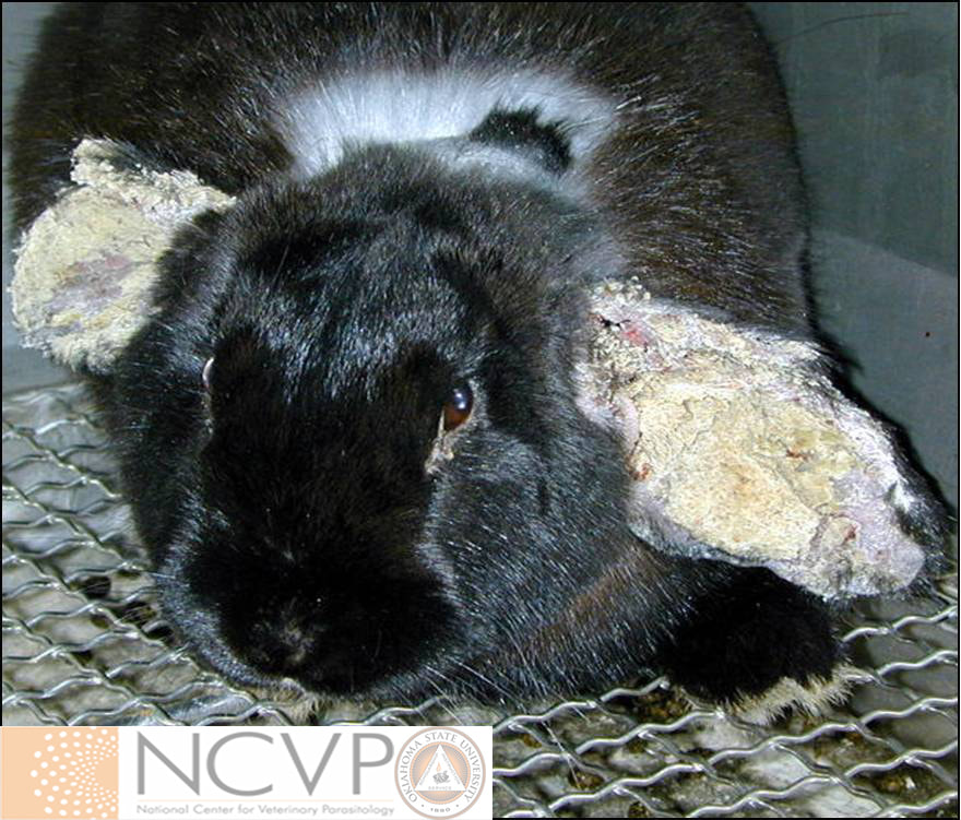
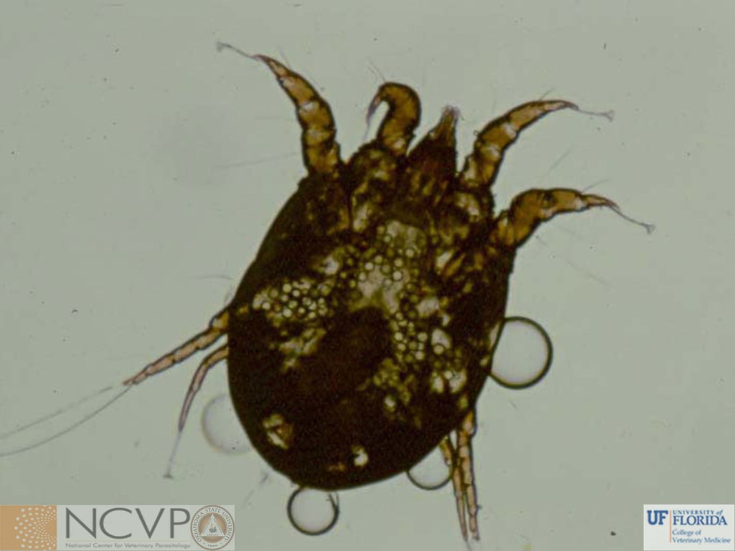
NCVP - National Center for Veterinary Parasitology

  • Home
  • Resources
    • People >
      • Directors and Advisory Board
      • Residents >
        • Boehringer Ingelheim Resident
        • Elanco Resident
        • IDEXX Resident
        • Merck Resident
        • Zoetis Resident
      • NCVP Alums
      • Clinical Parasitology Support
      • Additional Support
      • Sponsors
    • Parasite Image Database >
      • Protozoa
      • Arthropods
      • Nematodes
      • Trematodes
      • Cestodes
      • Acanthocephala
      • Tick-Borne Disease Agents
    • Case of the Month
    • Teaching and Research Materials >
      • Teaching Specimen Request
      • Parasite Jeopardy Games
      • Parasite Videos
    • Online Resources >
      • Parasitology Board Preparation
      • Additional Parasitology Resources
      • Resources for Educators
    • NCVP Board Member Resources
    • NCVP Zoom Backgrounds
    • Residents' Corner
  • News
    • Veterinary Parasitology in the News
    • NCVP Newsletter
  • Opportunities
    • Request for Proposals
    • Residency Application
    • Continuing Education in Parasitology
    • Positions Open in Veterinary Parasitology

Arthropods

Mites


​Cheyletiella spp.

Cheyletiella spp. are surface dwelling mites that are distinguished from other mites by their large palpal claws. Ventral view of a Cheyletiella sp. mite recovered from a cat. The large palpal claws and enlarged gnathosoma seen on the anterior portion are very characteristic of this mite. Cheyletiella spp. do not burrow, but do feed on the lymph and tissue fluids.
An egg of Cheyletiella. All developmental stages of Cheyletiella occur on the host. Eggs are glued to hairs loosely and may be ingested by the host and passed in the feces. Though this mite may live off of the host for up to 10 days, transmission is usually through direct contact between parent and offspring.

Chorioptes spp.

Chorioptes spp. infest a variety of animals including cattle, horses, and rabbits.​
Infestation with Chorioptes bovis on dairy cattle may result in a mild dermatitis on the back of the udder. Other locations where lesions commonly develop include the tailhead, feet, and legs.
Leg of a female Chorioptes. The cause of chorioptic mange in several species of domestic mammals, Chorioptes spp. have unjointed pedicels extending from their leg to tarsal suckers. These suckers occur on all of the legs on the male, and on leg pairs 1, 2, and 4 on the female.
Ventral aspect of a male Chorioptes bovis. Chorioptes bovis may infest horses causing foot mange, an intensely pruritic condition to which horses with long hair on their fetlocks are particularly susceptible.​

Demodex spp.

Demodex sp. mite ​
Demodex spp. are elongated mites found in sebaceous glands and hair follicles of their respective hosts. Transmission is generally direct from dam to offspring during the neonatal period.
Demodex canis
Demodex canis found in the skin scraping of a dog. Demodex specimens may also be seen on fecal flotation if pruritic animals ingest the mites while grooming.
Demodex caprae
in a skin scraping from a goat with severe demodicosis. Goats with demodicosis present with multiple nodular skin lesions on the face, neck, axillary region, and/or udder.​
Demodex cati
Lateral view of Demodex cati recovered from the ear canal of a domestic cat. Though usually non-pathogenic and considered natural flora of cats, immune suppression may lead to overpopulation of this mite. The crusting, pruritic lesions associated with D. cati generally involves the pinnae, eyelids, head, and neck. ​
Demodex gatoi
Demodex gatoi, another common Demodex sp. mite on cats, is found more superficially than Demodex cati and is often removed during grooming and thus not recovered on skin scraping. Instead, this mite is most commonly found in fecal flotations.

Dermanyssus spp.

Dermanyssus mites are frequently found on both domestic and wild birds. These mites occasionally invade dwellings and bite people and pets; the source is usually an abandoned nest nearby.

Eutrombicula spp.

Classified as a chigger mite, Eutrombicula may infest domestic mammals and produce a syndrome referred to as trombiculosis, characterized by pruritic skin papules or crusty eruptions.

Harpirhynchus

Ventral view of Harpirhynchus, a mite of the brown cowbird (Molothrus ater). Little is known about these mites, but they seem to burrow beneath the cowbird's skin and form cysts.​
Cysts on a brown cowbird formed by Harpirhynchus sp. The cysts are filled with mites, keratin, and cellular debris.​

Knemidokoptes spp.

Knemidokoptes, also known as the scaly leg mite, burrow under the scales of the non-feathered areas of birds. Infestations can lead to disfigurement and death of the birds.
Ventral and dorsal view of Knemidokoptes mutans. To collect and diagnose infestation of this mite, skin scrapings should be taken from the periphery of lesions.
A budgie infested with Knemidokoptes, with characteristic scaly proliferative lesions on the non-feathered portions of the face. The burrowing of the mite causes the integument to exude a serous fluid that hardens into a crust. Birds acquire the mites through direct contact with other infested birds or with contaminated fomites.

Notoedres cati

Ventral aspect of Notoedres cati collected from a cat. Notoedres can be differentiated from Sarcoptes by the presence of a dorsal rather than a terminal anus, and prominent rounded scales on its dorsum as opposed to the pointed scales found on Sarcoptes sp.
Lesions of Notoedres cati in a rabbit. Although usually found on cats, this mite may also infest rabbits. Infestations may begin on the ears and back of the neck and extend to the face and rest of the body.

Ophionyssus natricis

Ventral view of Ophionyssus natricis, a blood feeding mite of captive reptiles. These mites may be found under the scales of several species of snakes and lizards, notably around the rim of the eye. Heavy infestations lead to anemia, presenting as listlessness, and may lead to death.

Oribatid Mite

Scheloribates laevigatus
An oribatid mite, Scheloribates laevigatus, is considered the most efficient intermediate host for Moniezia throughout the world.

Ornithonyssus spp.

​Ornithonyssus mites infest birds in temperate, tropical, and subtropical areas.
Ornithonyssus sylviarum
Ornithonyssus sylviarum, known as the northern fowl mite, on the head of a chicken. Found in temperate regions worldwide, this mite can be seen on the skin among feathers, often concentrating around the vent, causing a dark discoloration.

Otodectes cynotis

Otodectes cynotis infests the ear canals of primarily dogs and cats.

Psorobia ​sp.

Dorsal view of a species of Psorobia. The most economically important member of the Psorergatidae family is Psorobia ovis. Known as the sheep itch mite, Psorobia ovis infests domestic sheep, causing pruritus, wool loss, dry scruffy skin, and erythema. Merinos are considered to be especially susceptible. Similar to Demodex, Psorobia mites live in hair follicles

Psoroptes spp.

Infestation of Psoroptes cuniculi in the ears of rabbits can produce a large amount of serum and hyperkeratotic crusts inside the ears.
Adult female Psoroptes cuniculi collected from a goat ear swab. Psoroptes cuniculi may be found on rabbits and ruminants. Other species of the same genus may be found on horses and wildlife.
Crusted lesions from Psoroptes cuniculi infestation in a goat. Lesions consist of exudative dermatitis and hair loss, often beginning in the ear and spreading to the head and neck. The mites live superficially on the skin surface and are easily collected with a superficial skin scraping or scabs that have been broken apart.
Lesions from Psoroptes ovis on the ventrum of a sheep. Once of considerable economic importance as the cause of psoroptic mange in ruminants, Psoroptes ovis has been eradicated from domestic sheep in many areas of the world.

Raillietia auris

Dorsal view of Raillietia auris. This mite lives in the ear canals of domestic ruminants and has been associated with Mycoplasma sp. and otitis externa.

Sarcoptes scabiei

Sarcoptes scabiei is a small, round mite that burrows in the skin of domestic and wild animals. Note the long, unjointed pedicels extending from the legs, terminating in suckers.
Infestations with Sarcoptes scabiei can produce a severely pruritic, erythematic dermatitis with alopecia, hyperkeratosis, and crust formation.
The intense pruritus experienced with scabies is due to the mite burrowing into the dermis to lay eggs, molt, and feed on young epidermal cells and lymph. In some animals, lesions severely encrusted with coagulated exudate may occur with or without pruritus, a condition known as crusted scabies. ​

Trixacarus caviae

The sarcoptiform mite of guinea pigs. Clinical cases present with widespread alopecia, intense pruritis, keratization, and occasionally seizures. A key identifying feature of this mite is the long, unsegmented pedicels on leg pairs 1 and 2 terminating in suckers.

    Categories

    All
    Bugs
    Crustaceans
    Fleas
    Flies
    Lice
    Mites
    Ticks