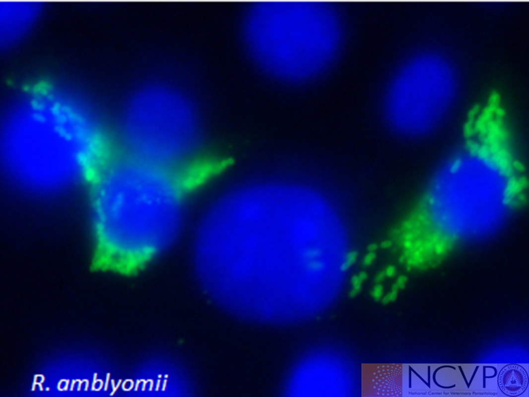
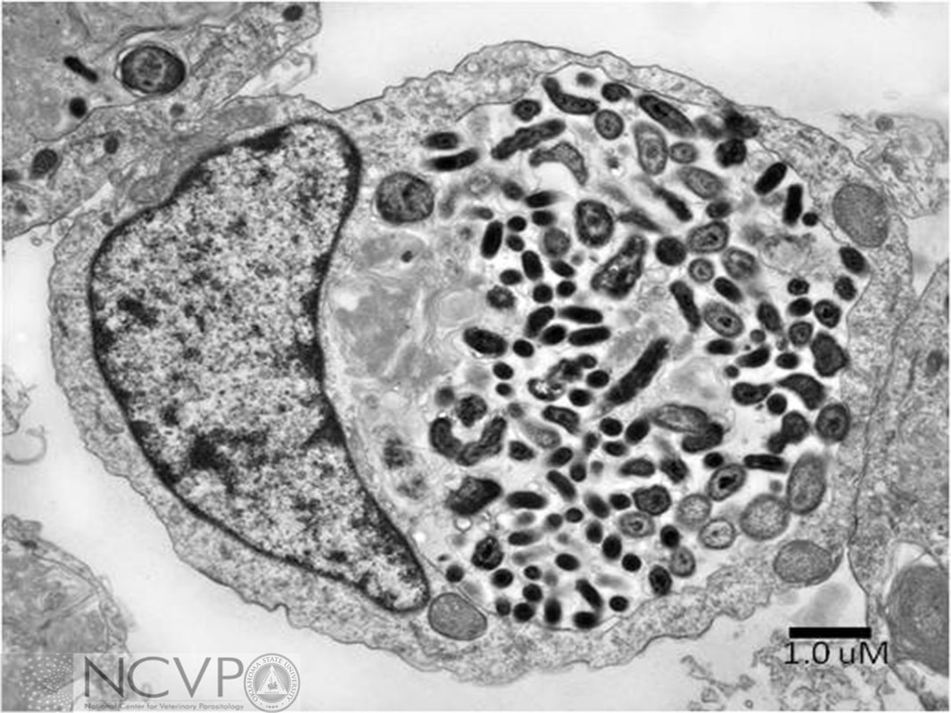
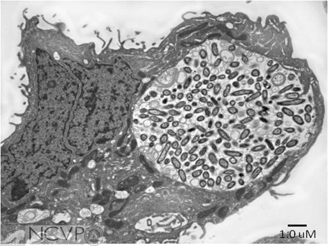
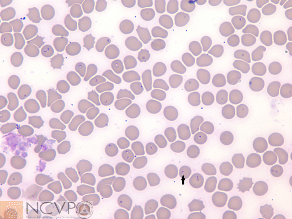
NCVP - National Center for Veterinary Parasitology

  • Home
  • Resources
    • People >
      • Directors and Advisory Board
      • Residents >
        • Boehringer Ingelheim Resident
        • Elanco Resident
        • IDEXX Resident
        • Merck Resident
        • Zoetis Resident
      • NCVP Alums
      • Clinical Parasitology Support
      • Additional Support
      • Sponsors
    • Parasite Image Database >
      • Protozoa
      • Arthropods
      • Nematodes
      • Trematodes
      • Cestodes
      • Acanthocephala
      • Tick-Borne Disease Agents
    • Case of the Month
    • Teaching and Research Materials >
      • Teaching Specimen Request
      • Parasite Jeopardy Games
      • Parasite Videos
    • Online Resources >
      • Parasitology Board Preparation
      • Additional Parasitology Resources
      • Resources for Educators
    • NCVP Board Member Resources
    • NCVP Zoom Backgrounds
    • Residents' Corner
  • News
    • Veterinary Parasitology in the News
    • NCVP Newsletter
  • Opportunities
    • Request for Proposals
    • Residency Application
    • Continuing Education in Parasitology
    • Positions Open in Veterinary Parasitology

Tick-Borne Disease Agents

Rickettsia


Anaplasma marginale

Transmitted by Dermacentor spp. (Dermacentor andersoni and Dermacentor variabilis in the United States). Romanowsky stained bovine erythrocytes containing Anaplasma marginale, which are dense, homogeneously staining blue-purple inclusions 0.3-1.0 µm in diameter that are typically located toward the margins of infected cells.

Anaplasma phagocytophilum

Transmitted by Ixodes spp. (Ixodes pacificus and Ixodes scapularis in the United States). Romanowsky stained canine blood smear containing morulae (arrow) of Anaplasma phagocytophilum within a neutrophil. Infections with Anaplasma phagocytophilum are most common in the northeastern United States, in the upper Midwest, and along the West Coast.

Anaplasma platys

Transmistted by Rhipicephalus sanguineus in other parts of the world, which is the suspected vector in the United States. Romanowsky stained canine blood film containing Anaplasma platys (PCR confirmed), which are intraplatelet organisms within vacuoles.

Ehrlichia canis

Transmitted by Rhipicephalus sanguineus. Romanowsky stained canine blood smear containing morulae (arrow) of Ehrlichia canis within a circulating monocyte. Infections with Ehrlichia canis can result in severe clinical disease in dogs (canine monocytic ehrlichiosis) and seem to occur most commonly in the southern United States, including areas of Arizona, southern California, Louisiana, Oklahoma and Texas. Ehrlichia chaffeensis is another tick-borne rickettia infecting monocytes that appears morphologically very similar to Ehrlichia canis in blood films. However, Ehrlichia chaffeensis is transmitted by Amblyomma americanum ticks. In dogs, Ehrlichia chaffeensis infections are rarely clinical. In humans, however, ehrlichiosis caused by Ehrlichia ​chaffeensis is considered the most common tick-borne disease in the southern United States.​

Ehrlichia ewingii

Transmitted by Amblyomma americanum. Romanowsky stained canine blood smear containing morulae (arrow) of Ehrlichia ewingii within a neutrophil. Infections with Ehrlichia ewingii tend to be clinically milder than with Ehrlichia canis, and are most commonly seen in the middle southern United States.

Rickettsia spp.

Spotted fever group Rickettsia spp. are transmitted by a number of different tick vectors, including Amblyomma americanum, Amblyomma maculatum, Dermacentor spp., and Rhipicephalus sanguineus, and are obligately intracellular organisms. Photo kindly provided by Dr. Ed Shaw, Oklahoma State University.

Coxiella burnetii (no longer considered a rickettsia)
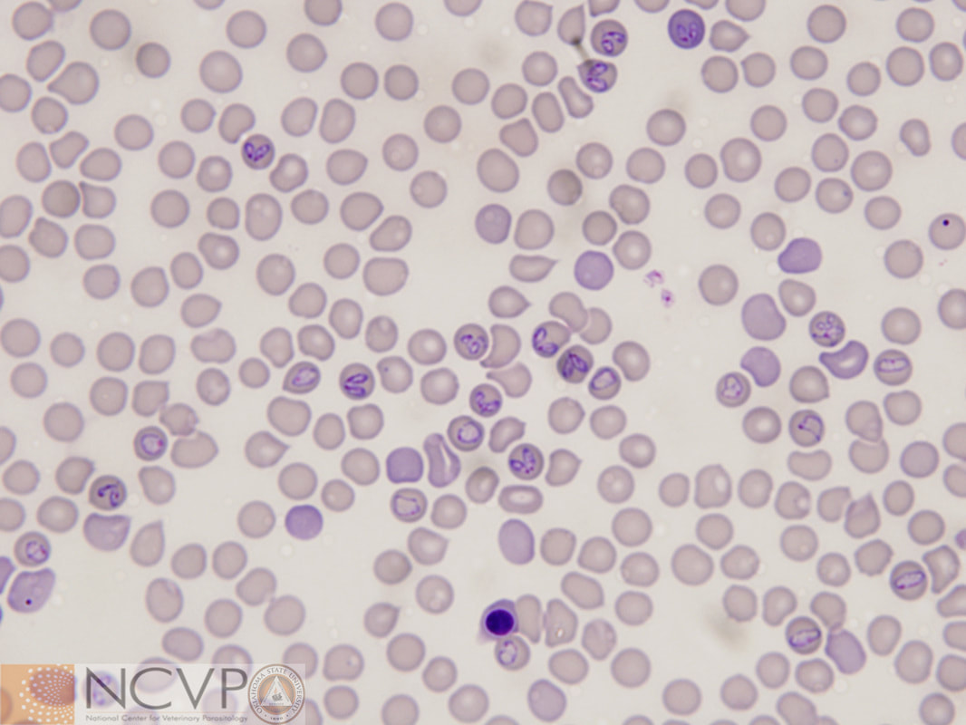
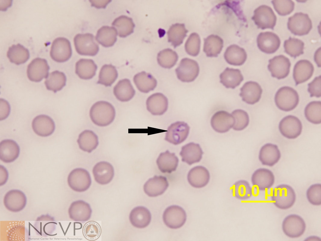
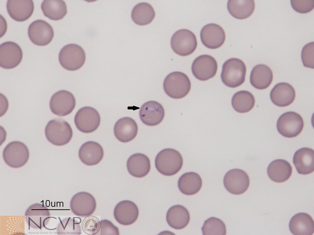
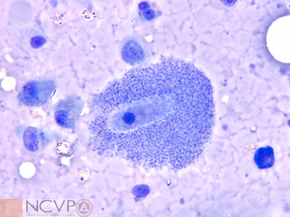
Electron micrograph of Coxiella burnetii within an IDE8 cell. Photo kindly provided by Dr. Ed Shaw, Oklahoma State University.
Electron micrograph of Coxiella burnetii within a VERO cell. Photo kindly provided by Dr. Ed Shaw, Oklahoma State University.

Protozoa


Babesia canis

Transmitted by various tick species, depending on the subspecies of Babesia canis. Piroplasms of Babesia canis in an erythrocyte on a Romanowsky stained blood film from an infected dog. Piroplasms of Babesia canis are 4-5 µm, pear-shaped, and typically paired. Piroplasms are the infective stage to feeding ticks.

Babesia bigemina

Transmitted by Rhipicephalus (Boophilus) spp. Piroplasms of Babesia bigemina in erythrocyte on Romanowsky stained blood smear from an infected cow. Piroplasms of Babesia bigemina are 4.5 x 2.5 µm, teardrop-shaped, and typically paired. Piroplasms are the infective stage to feeding ticks.

Babesia gibsoni

A tick vector has not been definitively demonstrated in the United States. Piroplasm of Babesia gibsoni in an erythrocyte on a Romanowsky stained blood film from an infected dog. Piroplasms of Babesia gibsoni are 1-3 µm and signet ring shaped. In other parts of the world, Babesia gibsoni is transmitted by Rhipicephalus sanguineus and Haemaphysalis species. In the Unites States, Rhipicephalus sanguineus is a suspected tick vector, but most Babesia gibsoni transmission is thought to occur through dog fighting.

Cytauxzoon felis

Transmitted by Amblyomma americanum and Dermacentor variabilis. Macrophage containing Cytauxzoon felis schizonts. Romanowsky stained lung impression smear.​
Romanowsky stained blood film with numerous erythrocytes containing Cytauxzoon felis piroplasms. Piroplasms are the infective stage to feeding ticks. Amblyomma americanum is thought to be the primary natural tick definitive host and vector.

Hepatozoon americanum

Transmitted by Amblyomma maculatum. Characteristic "onion skin" cyst of Hepatozoon americanum in a histologic muscle preparation from an infected dog. These cysts are associated with parasite schizogony within leukocytes situated between striated muscle fibers, and are pathognomonic for infection with this parasite. Here, the parasite has invaded the host cell and induced cyst formation, but has not yet begun to undergo schizogony.
Gamont of Hepatozoon americanum in a leukocyte on Romanowsky stained blood film from an infected dog. Gamonts are rarely seen on blood films, as parasitemia is typically extremely low. Gamonts are the infective stage to feeding ticks.

Theileria equi

Transmitted by Rhipicephalus, Hyalomma, and Dermacentor species. Romanowsky stained blood film with numerous erythrocytes containing Theileria equipiroplasms, the infective stage to feeding ticks. Equine piroplasmosis is endemic in parts of Asia, Africa, the Middle East, and Central and South America, but has been introduced into the United States on occasion.

    Categories

    All
    Protozoa
    Rickettsia