Home
Resources
People
>
Directors and Advisory Board
Residents
>
Boehringer Ingelheim Resident
Elanco Resident
IDEXX Resident
Merck Resident
Zoetis Resident
NCVP Alums
Clinical Parasitology Support
Additional Support
Sponsors
Parasite Image Database
>
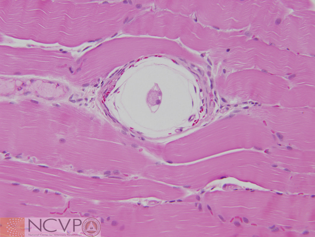
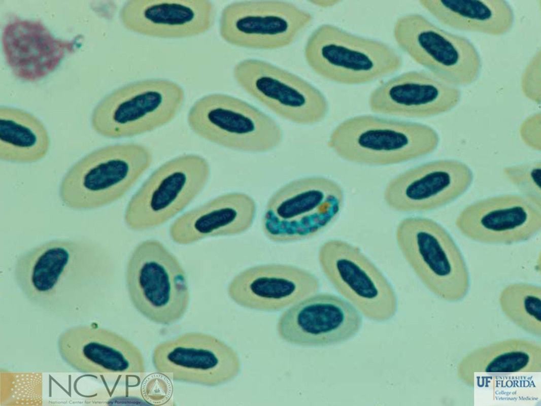
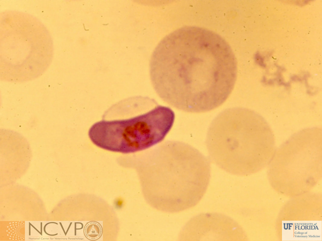
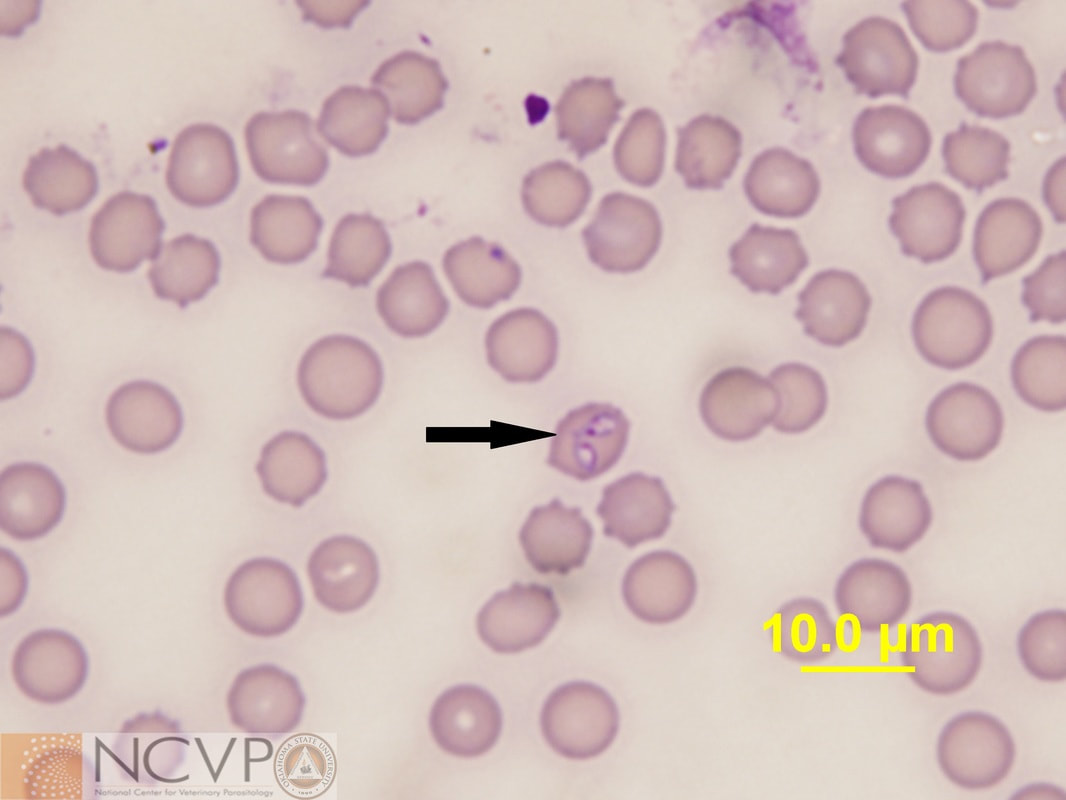
Protozoa
Arthropods
Nematodes
Trematodes
Cestodes
Acanthocephala
Tick-Borne Disease Agents
Case of the Month
Teaching and Research Materials
>
Teaching Specimen Request
Parasite Jeopardy Games
Parasite Videos
Online Resources
>
Parasitology Board Preparation
Additional Parasitology Resources
Resources for Educators
NCVP Board Member Resources
NCVP Zoom Backgrounds
News
Veterinary Parasitology in the News
NCVP Newsletter
Opportunities
Request for Proposals
Grant Portal
Residency Application
Continuing Education in Parasitology
Positions Open in Veterinary Parasitology
Home
Resources
People
>
Directors and Advisory Board
Residents
>
Boehringer Ingelheim Resident
Elanco Resident
IDEXX Resident
Merck Resident
Zoetis Resident
NCVP Alums
Clinical Parasitology Support
Additional Support
Sponsors
Parasite Image Database
>
Protozoa
Arthropods
Nematodes
Trematodes
Cestodes
Acanthocephala
Tick-Borne Disease Agents
Case of the Month
Teaching and Research Materials
>
Teaching Specimen Request
Parasite Jeopardy Games
Parasite Videos
Online Resources
>
Parasitology Board Preparation
Additional Parasitology Resources
Resources for Educators
NCVP Board Member Resources
NCVP Zoom Backgrounds
News
Veterinary Parasitology in the News
NCVP Newsletter
Opportunities
Request for Proposals
Grant Portal
Residency Application
Continuing Education in Parasitology
Positions Open in Veterinary Parasitology