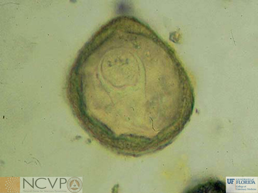
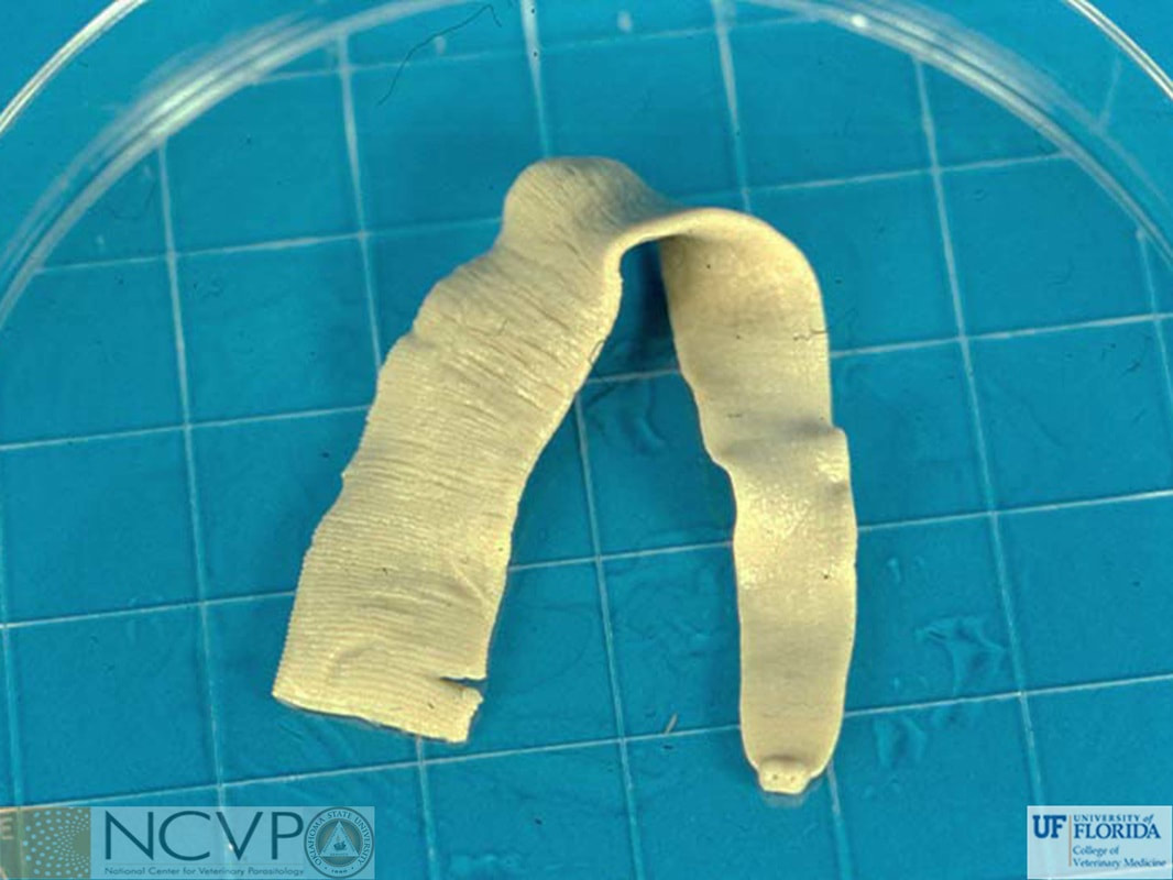
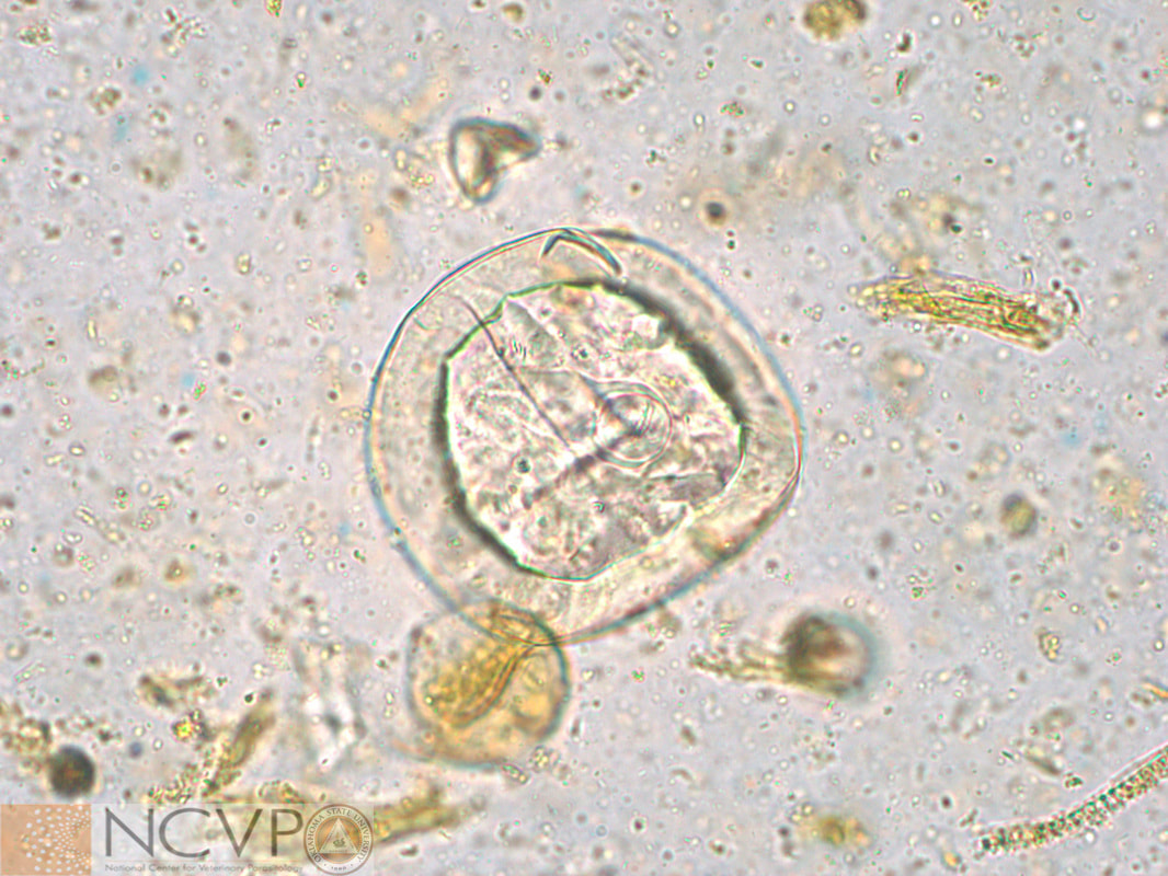
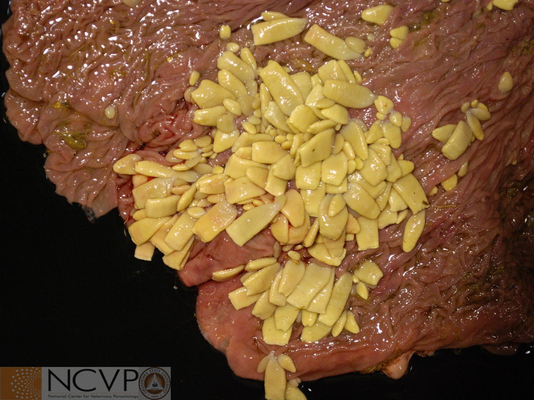
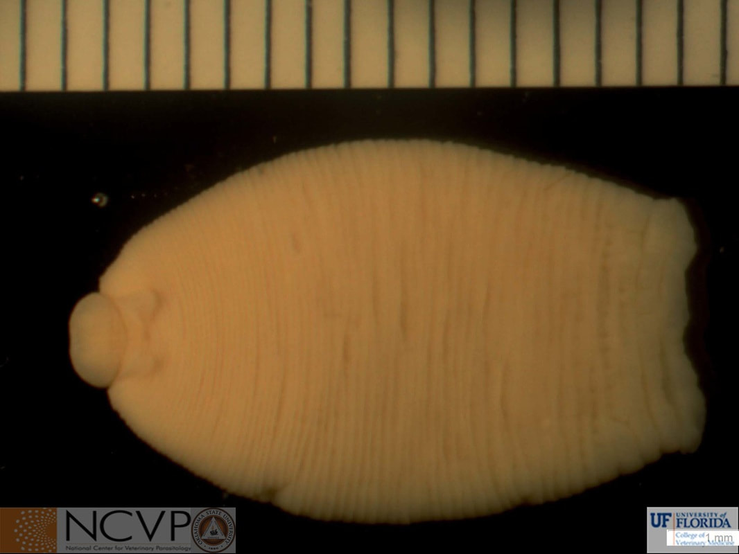
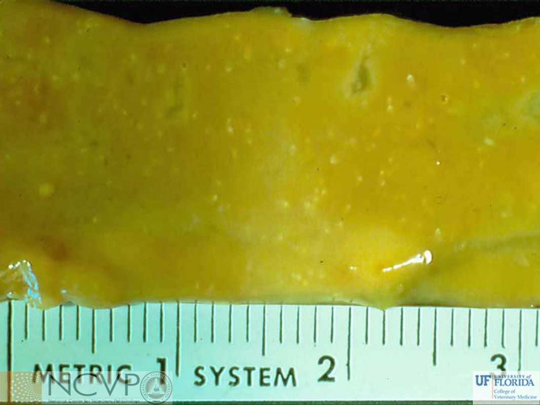
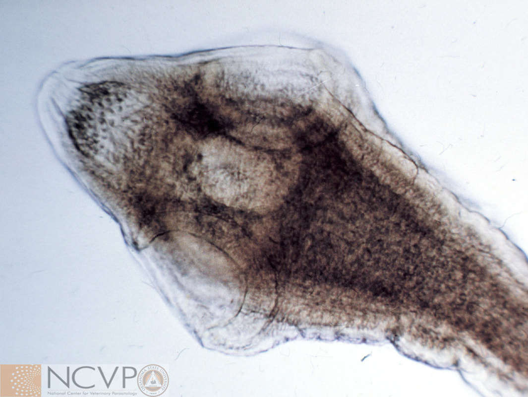
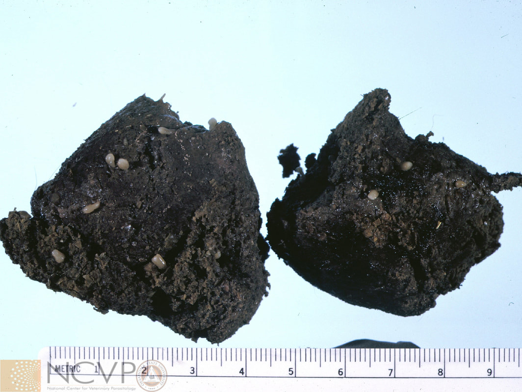
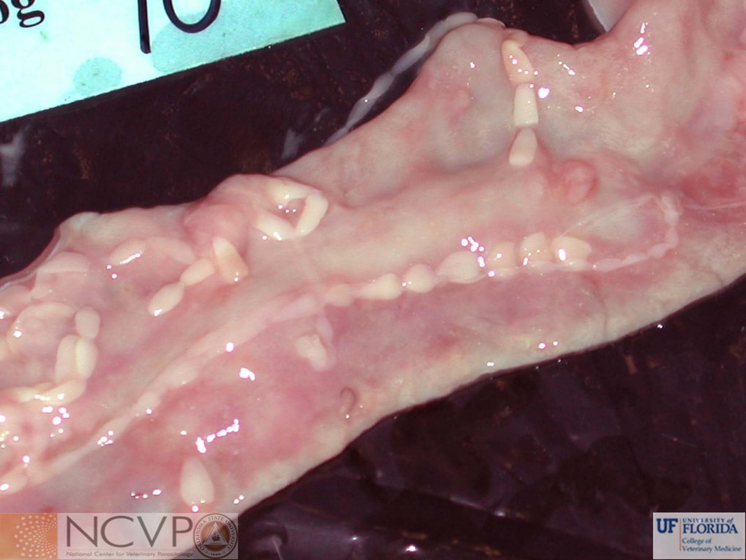
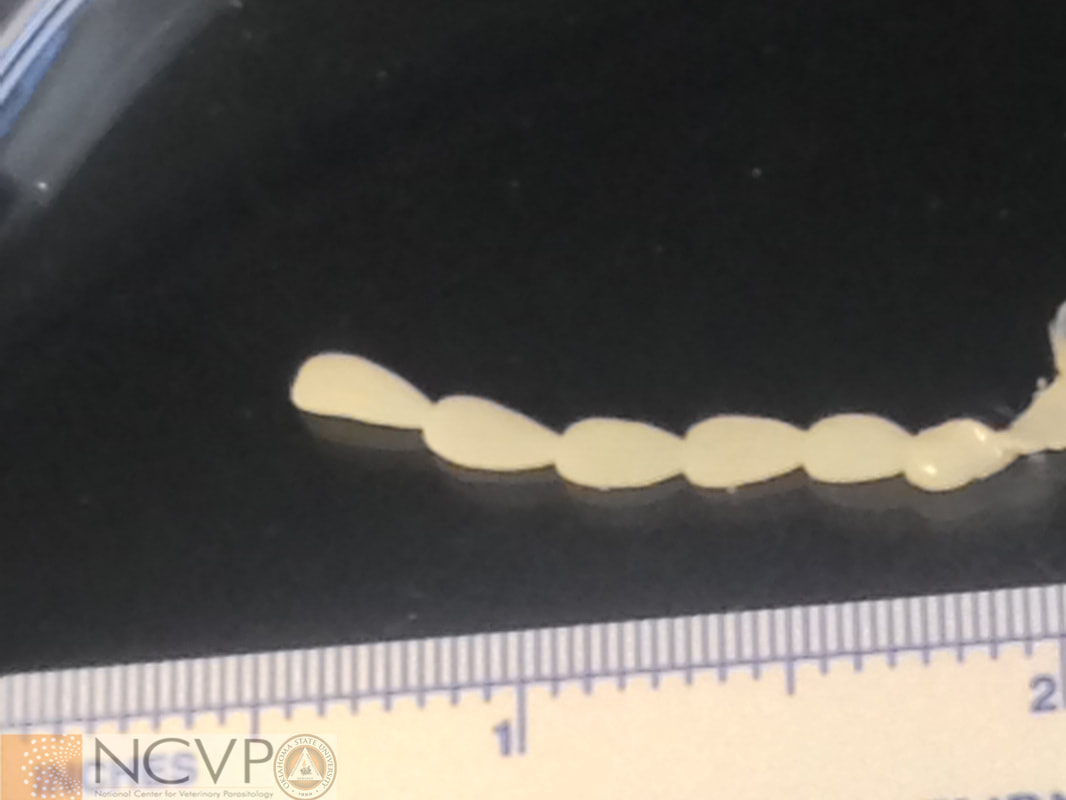
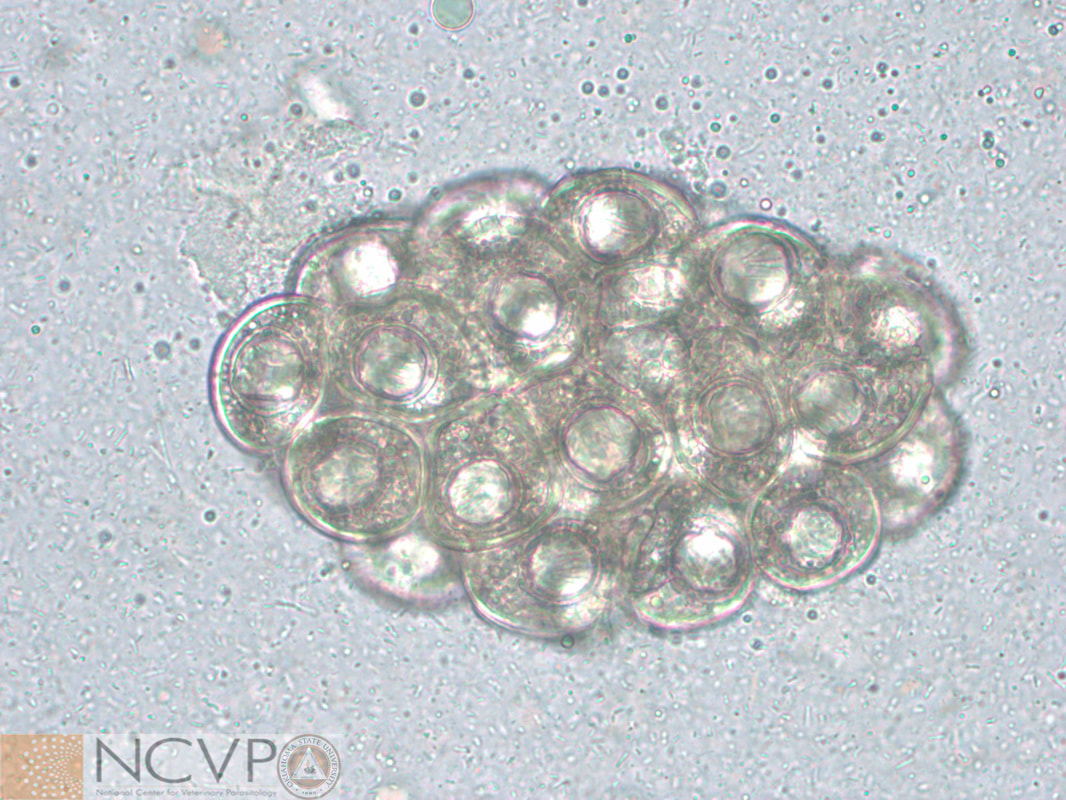
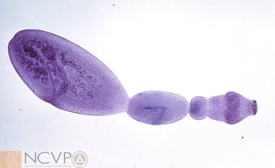
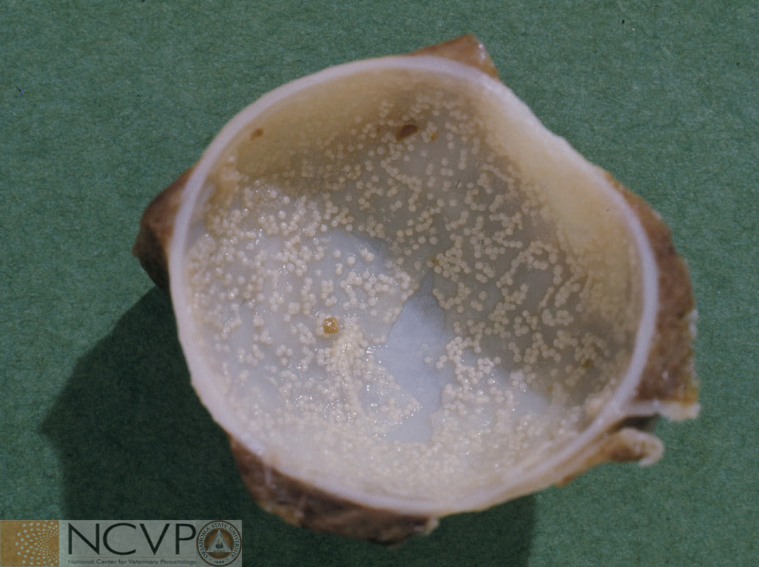
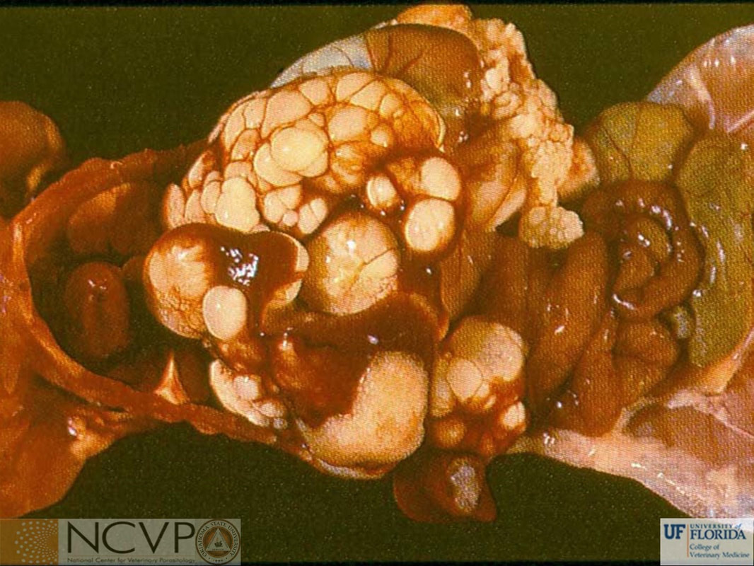
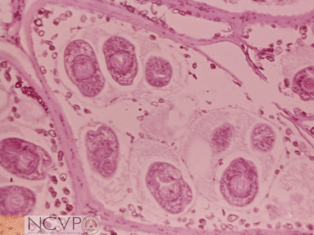
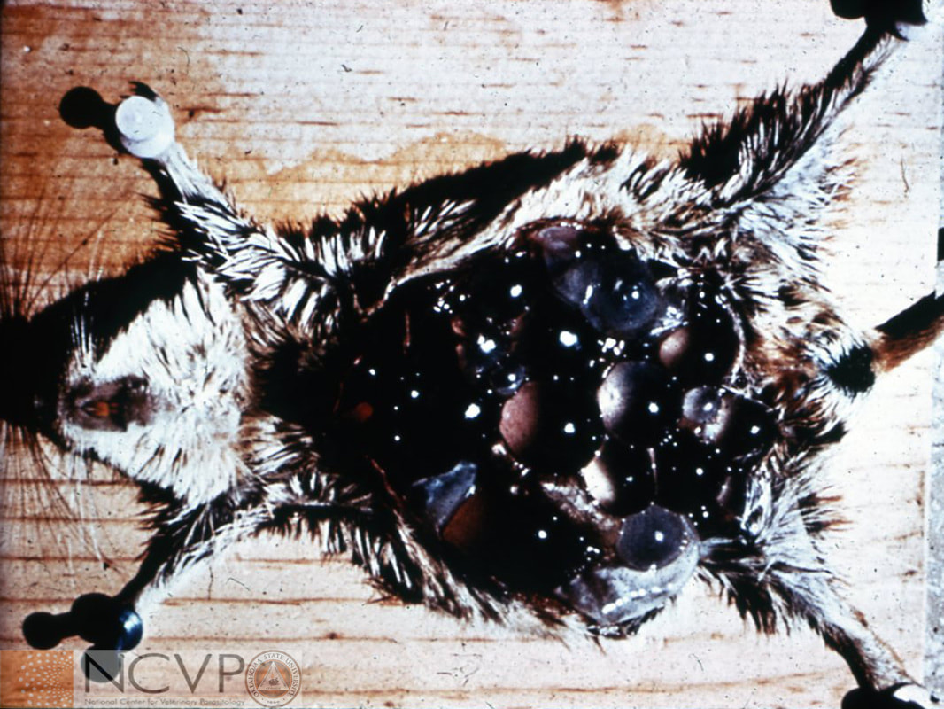
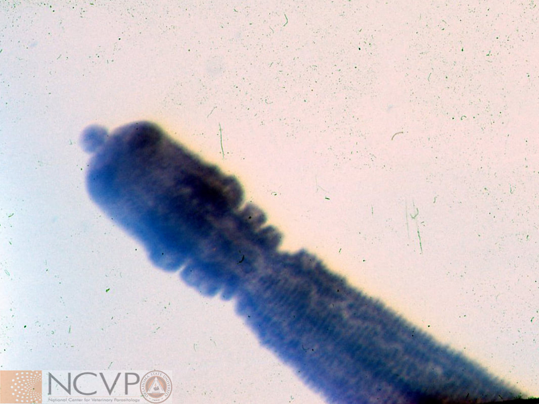
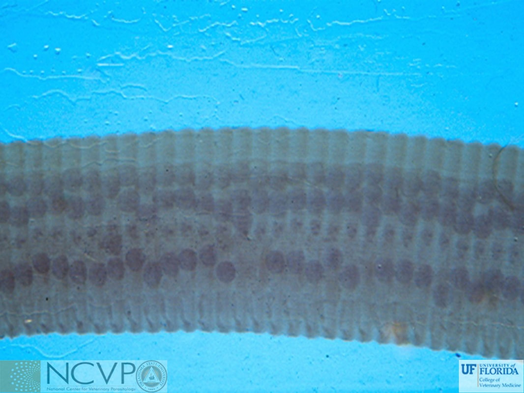
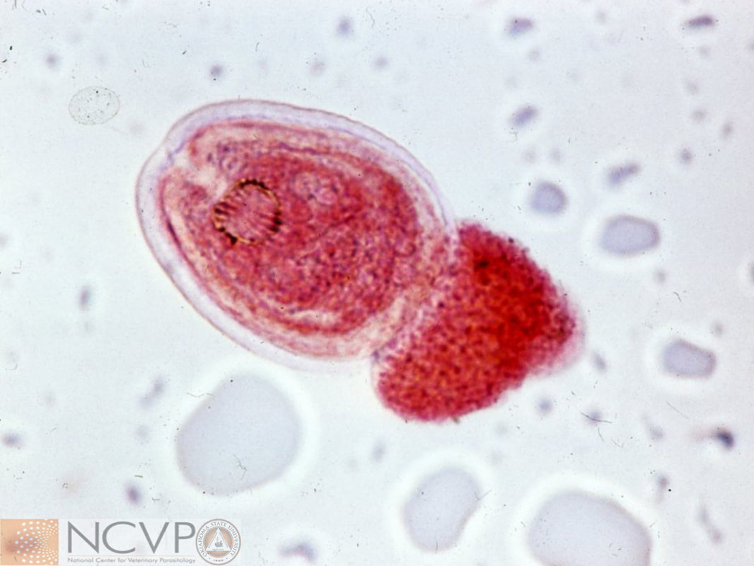
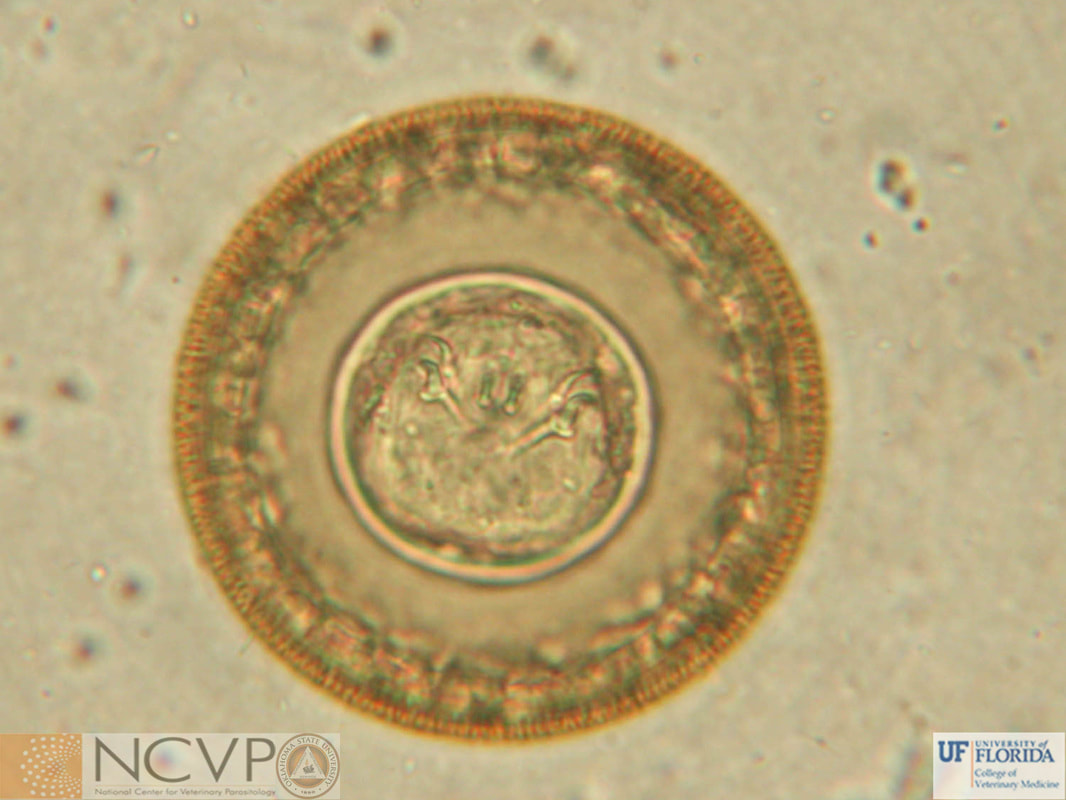
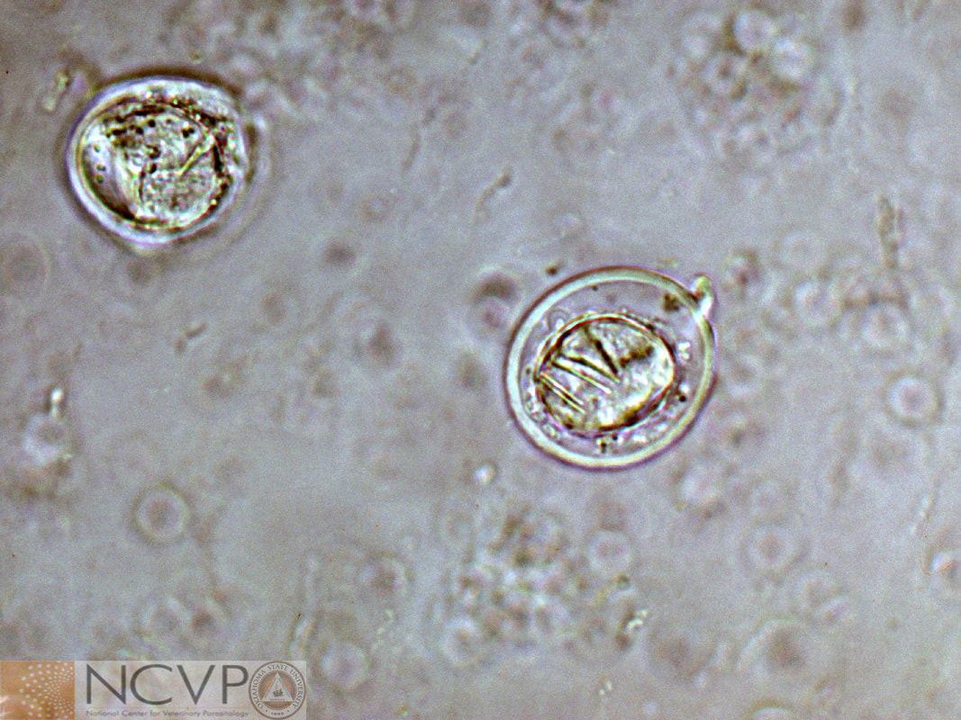
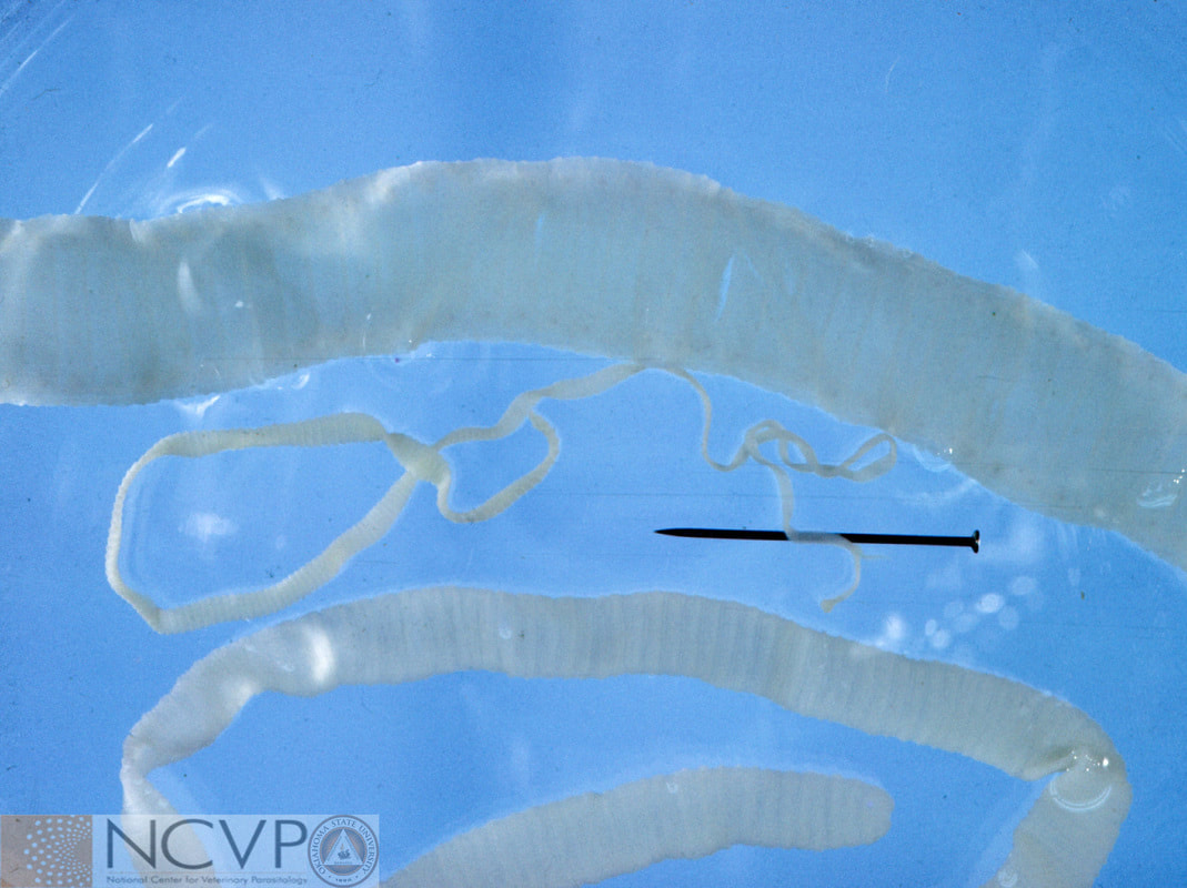
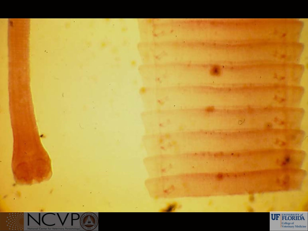
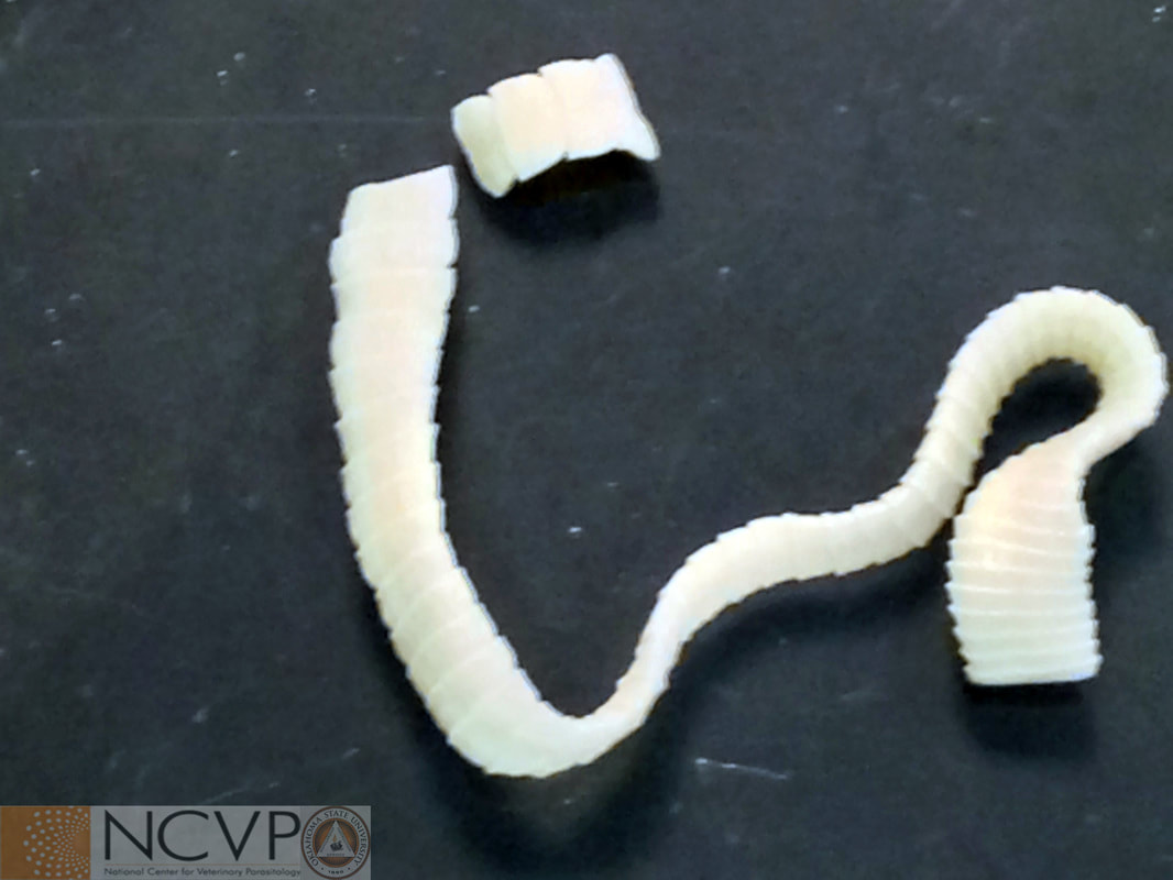
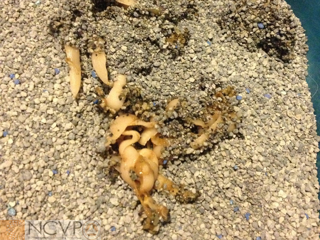
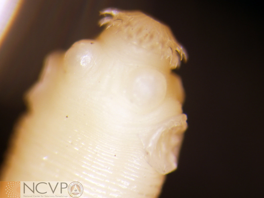
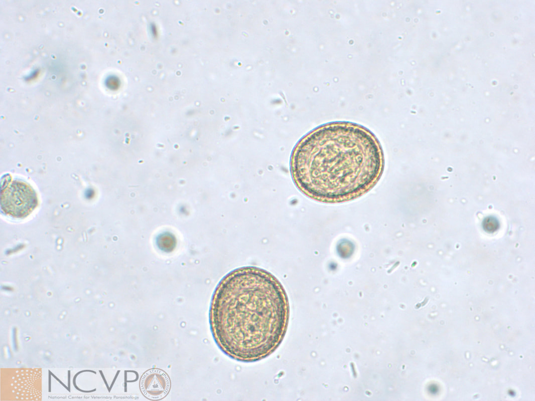
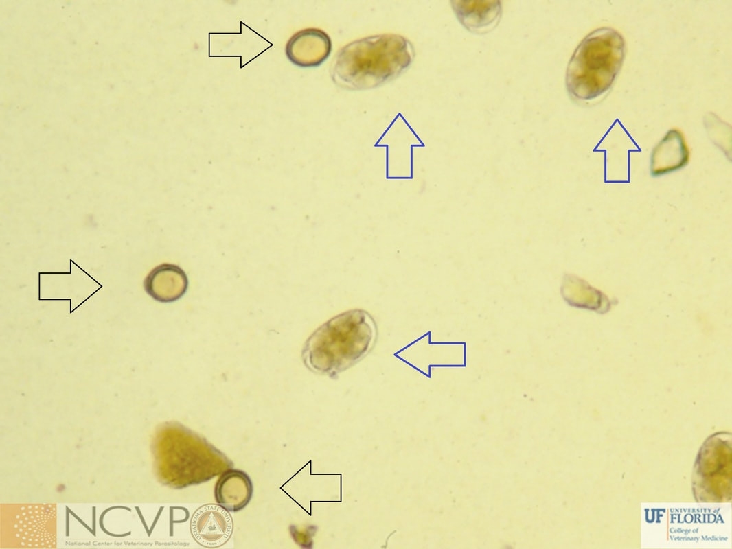
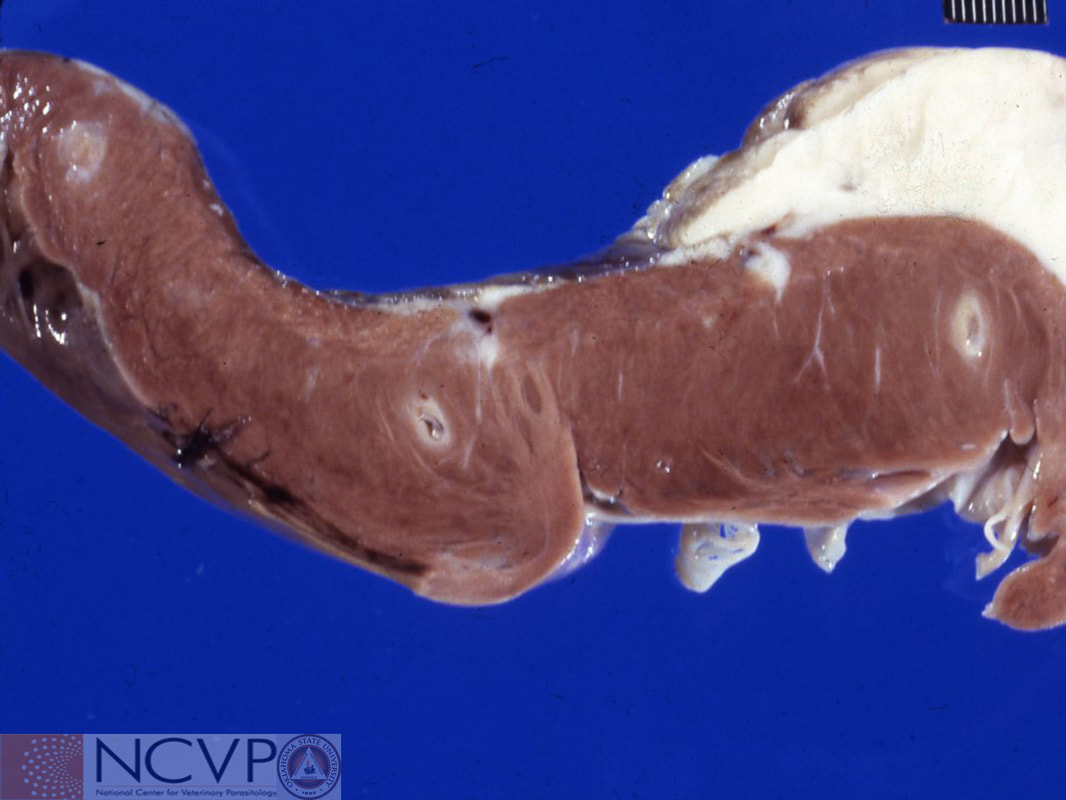
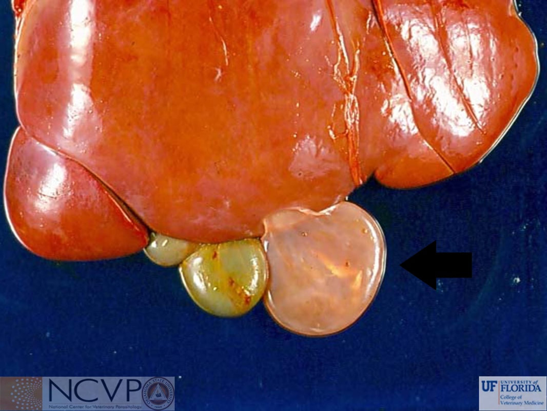
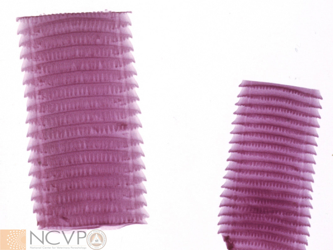
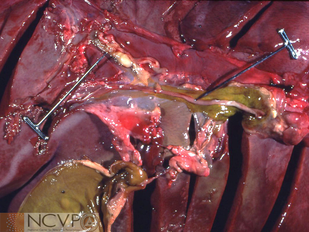
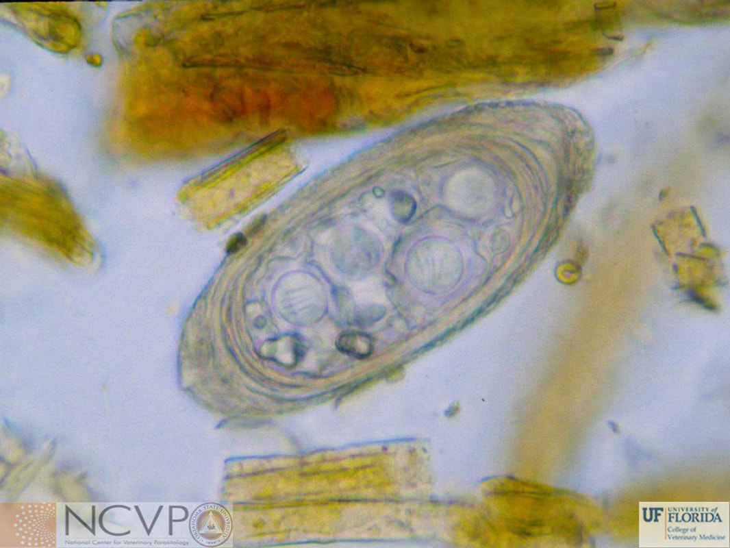
NCVP - National Center for Veterinary Parasitology

  • Home
  • Resources
    • People >
      • Directors and Advisory Board
      • Residents >
        • Boehringer Ingelheim Resident
        • Elanco Resident
        • IDEXX Resident
        • Merck Resident
        • Zoetis Resident
      • NCVP Alums
      • Clinical Parasitology Support
      • Additional Support
      • Sponsors
    • Parasite Image Database >
      • Protozoa
      • Arthropods
      • Nematodes
      • Trematodes
      • Cestodes
      • Acanthocephala
      • Tick-Borne Disease Agents
    • Case of the Month
    • Teaching and Research Materials >
      • Teaching Specimen Request
      • Parasite Jeopardy Games
      • Parasite Videos
    • Online Resources >
      • Parasitology Board Preparation
      • Additional Parasitology Resources
      • Resources for Educators
    • NCVP Board Member Resources
    • NCVP Zoom Backgrounds
    • Residents' Corner
  • News
    • Veterinary Parasitology in the News
    • NCVP Newsletter
  • Opportunities
    • Request for Proposals
    • Residency Application
    • Continuing Education in Parasitology
    • Positions Open in Veterinary Parasitology

Cestodes

Cyclophyllidean tapeworms


Anoplocephala magna

Eggs of Anoplocephala magna range 50-60 μm in diameter. They develop into a cysticercoid after ingestion by a free living oribatid mite, then develop into an adult once the infected mite is ingested by an equid.
Adult Anoplocephala magna. Note the lack of lappets on anterior end which differentiates them from Anoplocephala perfoliata. Adults may reach 80 cm in length and 2.5 cm wide

Anoplocephala perfoliata

Eggs of Anoplocephala perfoliata, a tapeworm of horses, are 65–80 μm in diameter, “D” shaped, and contain a distinct pyriform apparatus.
Adults of Anoplocephala perfoliata cluster at the ileocecal junction.
​Adult Anoplocephala perfoliata. Adults of this species are 5-8 cm in length and an average of 1.2 cm in width. It features lappets behind each sucker which aids in its identification.

Davainea proglottina

Cross section of adult Davainea proglottina, a tapeworm of gallinaceous birds worldwide. Adults are 0.5-3 mm in length and may be found in the duodenum.
Adult Davainea proglottina may be seen dotting this section of duodenum from a pigeon. Infection with this cestode occurs after a gallinaceous bird ingests a gastropod mollusk containing a cysticercoid.

Dipylidium caninum

​Here the rostellum of Dipylidium caninum is retracted.
​Proglottids of Dipylidium caninum are often readily apparent in canine feces
Adult Dipylidium caninum may reach 50 cm in length and infect canids, cats, and occasionally children. Infection is a result of ingestion of a flea containing a cysticercoid.
​Segments of the flea tapeworm (Dipylidium caninum) recovered from a dog. Note the characteristic cucumber-seed shape.
Eggs are found in clusters that are 120–200 μm in size. Individual eggs measure 35–60 μm in diameter and contain an embryo bearing hooks.

Echinococcus spp.

Adults of Echinococcus multilocularis and Echinococcus granulosus are only a few millimeters in length and consist of 3-5 segments
​On cut section, granular protoscolices are visible within this unilocular hydatid cyst of Echinococcus granulosus.
Echinococcus hydatid cysts
Hydatid cysts in a gerbil liver. Intermediate hosts become infected upon ingestion of eggs shed by canids.​
​Taeniid eggs of Taenia spp. and Echinococcus spp. are morphologically indistinguishable, measure 25-40 μm, and consist of a thick, striated wall surrounding a hexacanth embryo.

Echinococcus multilocularis

Infection of the intermediate host with Echinococcus multilocularis results in formation of the diffuse, multilocular cyst in the liver.
A mouse infected with Echinococcus multilocularis. The multilocular hydatid cysts are infective immature stages of Echinococcus multilocularis, and will establish infection once eaten by a canid.

Hymenolepis

Hymenolepis nana
The scolex of Hymenolepis nana, with four unarmed suckers and a retractable rostellum with a single circle of hooks. Known as the dwarf tapeworm, Hymenolepis nana infects the small intestines of rodents, simian primates, and humans.
Hymenolepis sp. ​
Mature proglottids of Hymenolepis. Adults are small to medium in size, with Hymenolepis nana being 25-40 mm long and Hymenolepis diminuta being 2-6 cm long.
Hymenolepis nana
Cysticercoid of Hymenolepis nana. In humans, Hymenolepis nana has a direct life cycle, with the cysticercoids developing in the villi of the small intestine, and auto infection is possible. In rodents, the life cycle may be either direct or indirect, with flour beetles or fleas serving as intermediate hosts.
Hymenolepis nana
Found on the fecal flotation of a desert rat, this egg of Hymenolepis nana measures 48x39 μm. The average is 44-62x30-55 μm. The eggs of this genus each have three delicate, enclosing membranes and three pairs of hooks.
Hymenolepis diminuta
Egg of Hymenolepis diminuta. This tapeworm is found in wild rodents and has been documented in humans and canids. The eggs of this genus each have three delicate, enclosing membranes and three pairs of hooks.

Mesocestoides spp.

​The eggs of Mesocestoides spp. are 30–40 μm in diameter and the embryonic hooks are readily visible.
​The small proglottids of Mesocestoides spp. are club-shaped and have a distinct parauterine organ containing eggs.
​The scolex of Mesocestoides spp. has four suckers, but no rostellum or hooks.

Moniezia spp.

Adult Moniezia spp. in ruminants often measure several meters in length. Infection is acquired through the ingestion of an oribatid mite containing a Moneizia cysticercoid.
Moniezia expansa
Scolex and mature proglottids of Moniezia expansa, the primary tapeworm of sheep. Adults of this species may reach a length of 600 cm. Their scolex has neither rostellum nor hooks and the segments are broader than long with two marginal genital pores.

Taenia spp.

This stained segment of an adult Taenia saginata shows the uterine branches and unilateral pore.
Unstained segments of a Taenia sp. adult.
Taenia taeniaeformis
Mass of Taenia taeniaeformis in cat litter expelled after a shelter cat was dewormed.
​The scolex of Taenia spp. is armed with two concentric rows of hooks, has four suckers (2 visible here), and is non-retractable.
Taeniid eggs of Taenia spp. and Echinococcus spp. are morphologically indistinguishable, measure 25-40 μm , and consist of a thick, striated wall surrounding a hexacanth embryo.
Taenia sp. and Ancylostoma caninum
Taenia eggs (black arrows) and Ancylostoma caninum (blue arrows) in a fecal flotation sample. Taenia eggs may not be seen on fecal flotation if still contained within tapeworm proglotids, making flotation a poor indicator of infection status.
Taenia saginata
Cysticecrosis (infection with intermediate stages of Taenia) in the heart of a bovine. Cattle act as an intermediate host and harbor the cysticerci within muscle tissue which, if not cooked properly, may infect humans, the definitive host, upon ingestion.
Taenia taeniaeformis
Strobilocercus being removed from a rat liver. Rodents serve as the intermediate host for Taenia taeniaeformis; the definitive host is domestic cats in the urban cycle and bobcats in the sylvatic cycle.
Taenia hydatigena
Taenia hydatigena cysticercus in a sheep liver. When an appropriate intermediate host (any wild or domestic ruminant) ingests the egg of Taenia hydatigena, the egg will hatch in the small intestine and the released oncosphere will reach the liver via the blood. The embryo then breaks out of the portal vessels and migrates through the liver, eventually leaving the liver as a cysticercus and attaching to the serosal surface of organs and mesentery to await ingestion by a canid.

Thysanosoma actinoides

Each segment of Thysanasoma actinoides bears a row of fringe posteriorly, allowing ready identification. Thysanasoma is exclusive to the western hemisphere, including mostly Mexico and the western USA. Adults measure 15-30 cm by 8 mm with a scolex of 1.5 mm wide.
​​Adult Thysanosoma in the hepatobilary tree of a sheep. Thysanosoma may inhabit the bile duct, pancreatic duct, or small intestine of not only sheep, but of cattle and deer as well.
Thysanosoma eggs are passed as packets, similar in size to the eggs of Haemonchus, containing 6-12 eggs. No pyriform apparatus is present, but hexacanth embryos may still be visible.

    Categories

    All
    Cyclophyllidean
    Pseudophyllidean