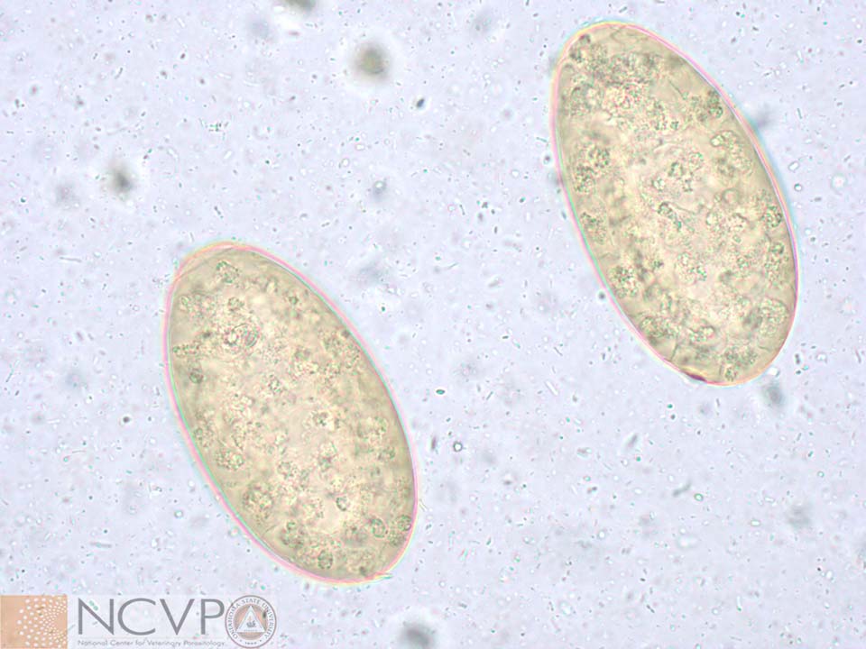
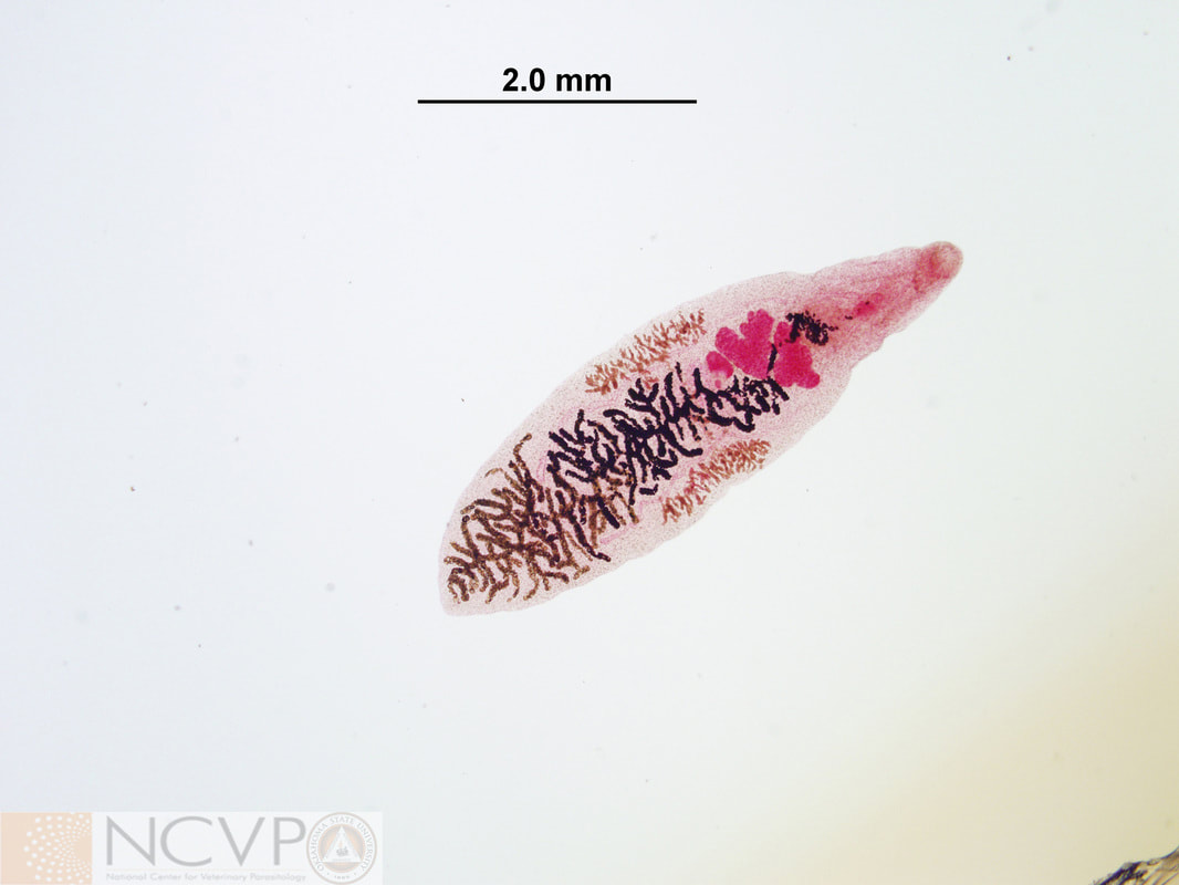
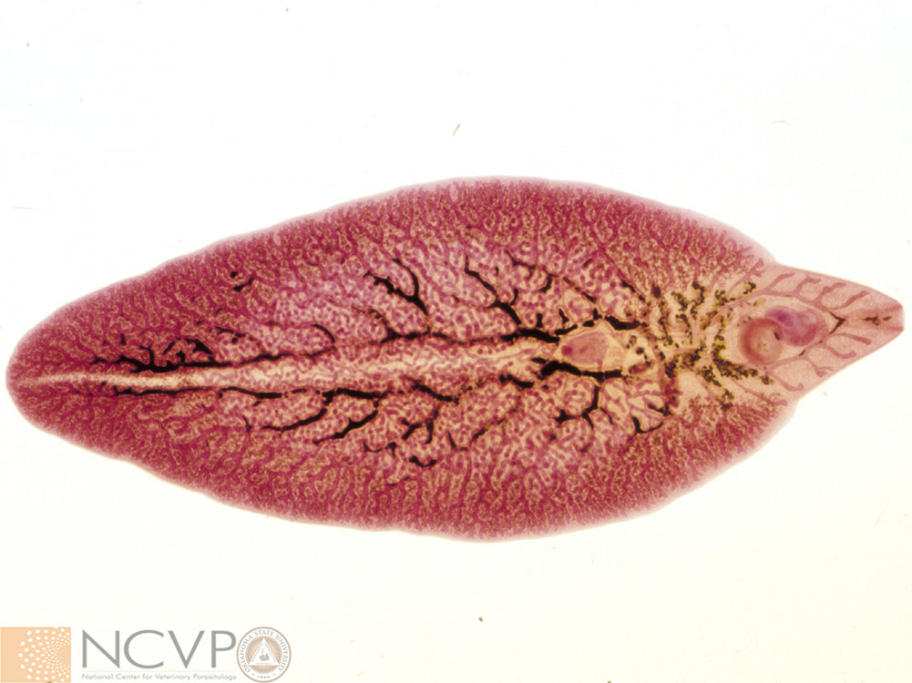
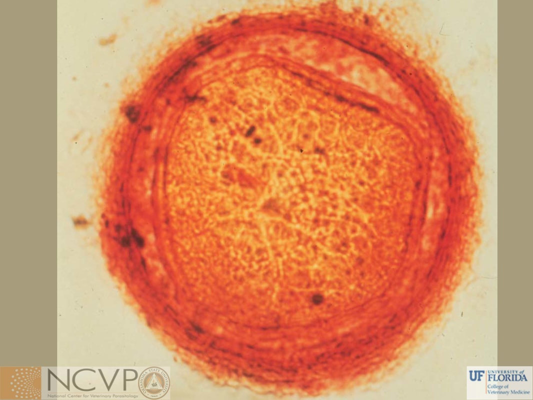
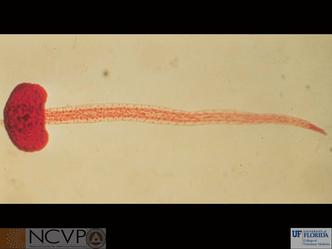
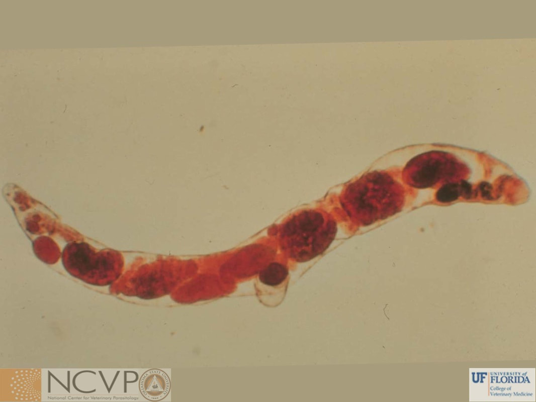
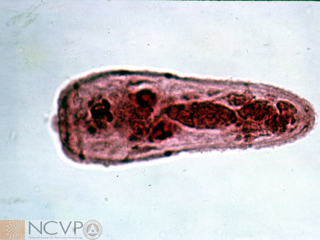
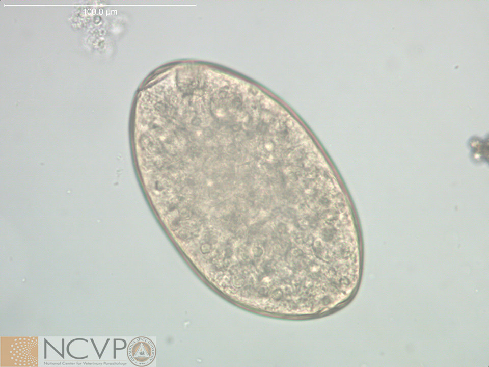
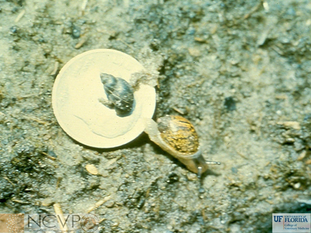
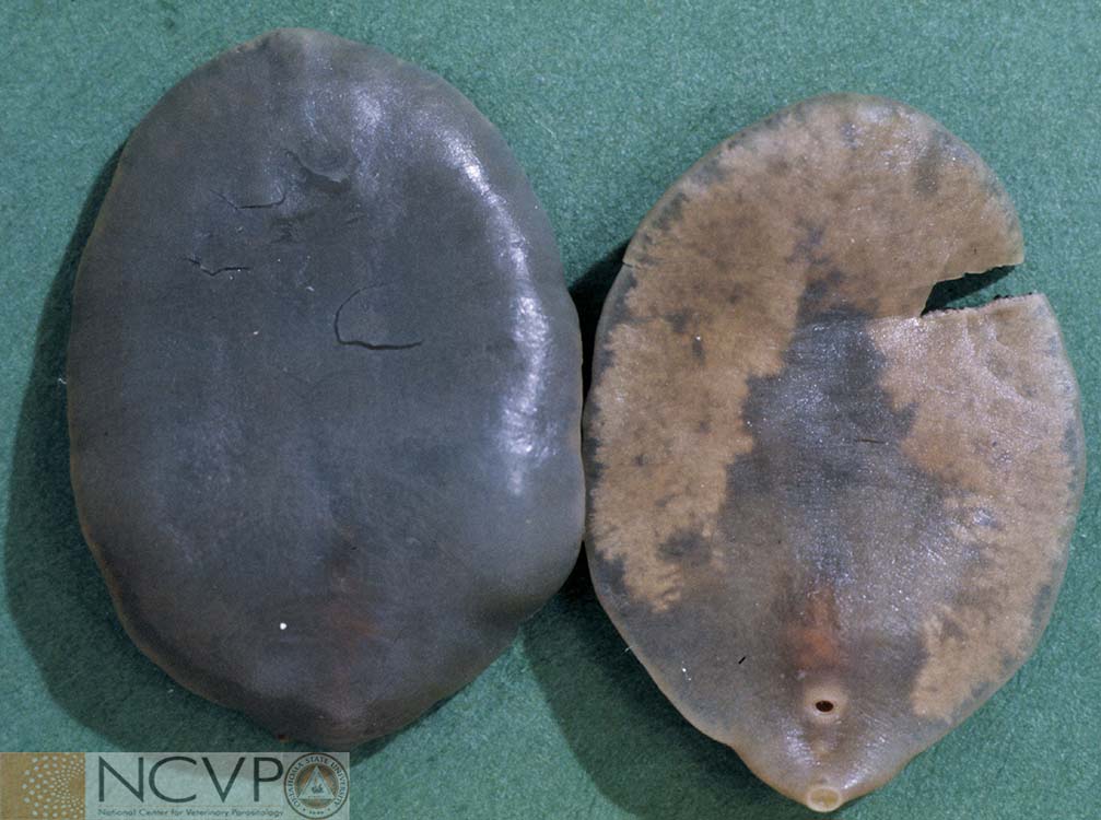
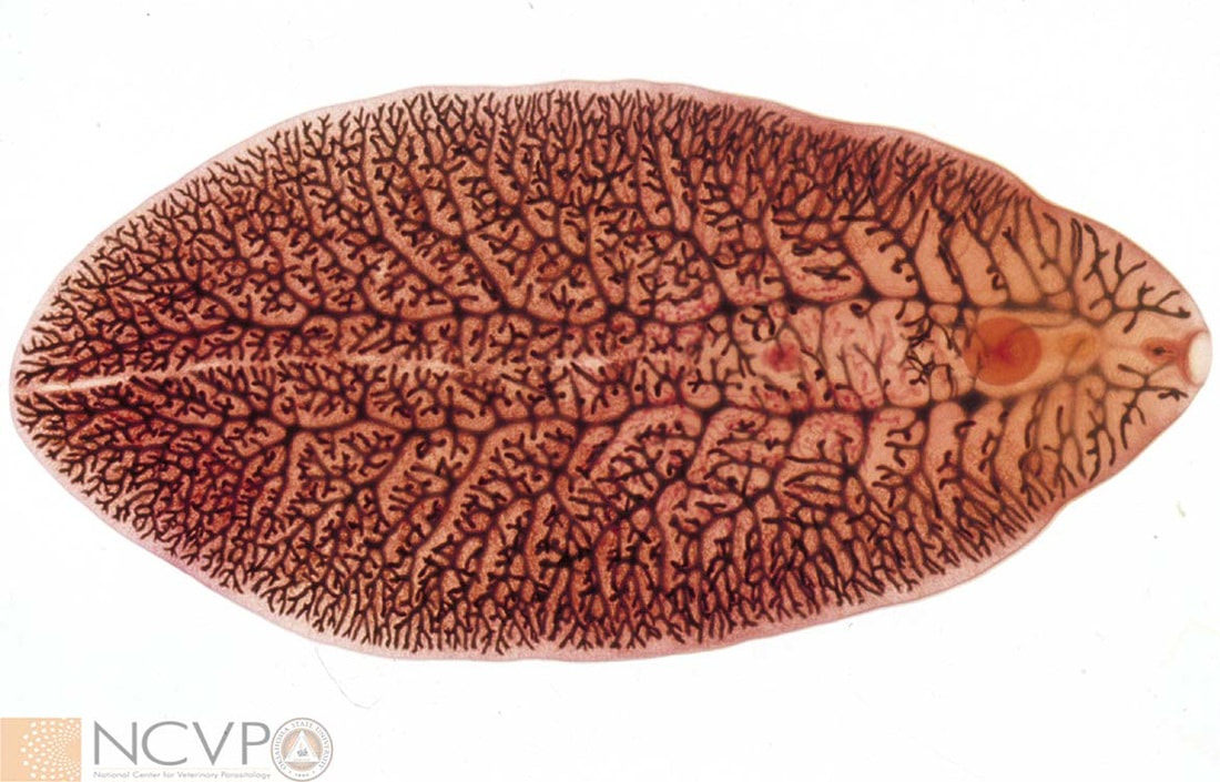
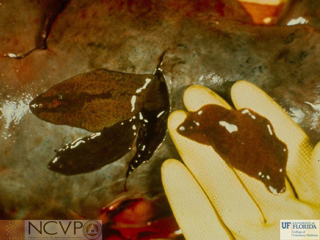
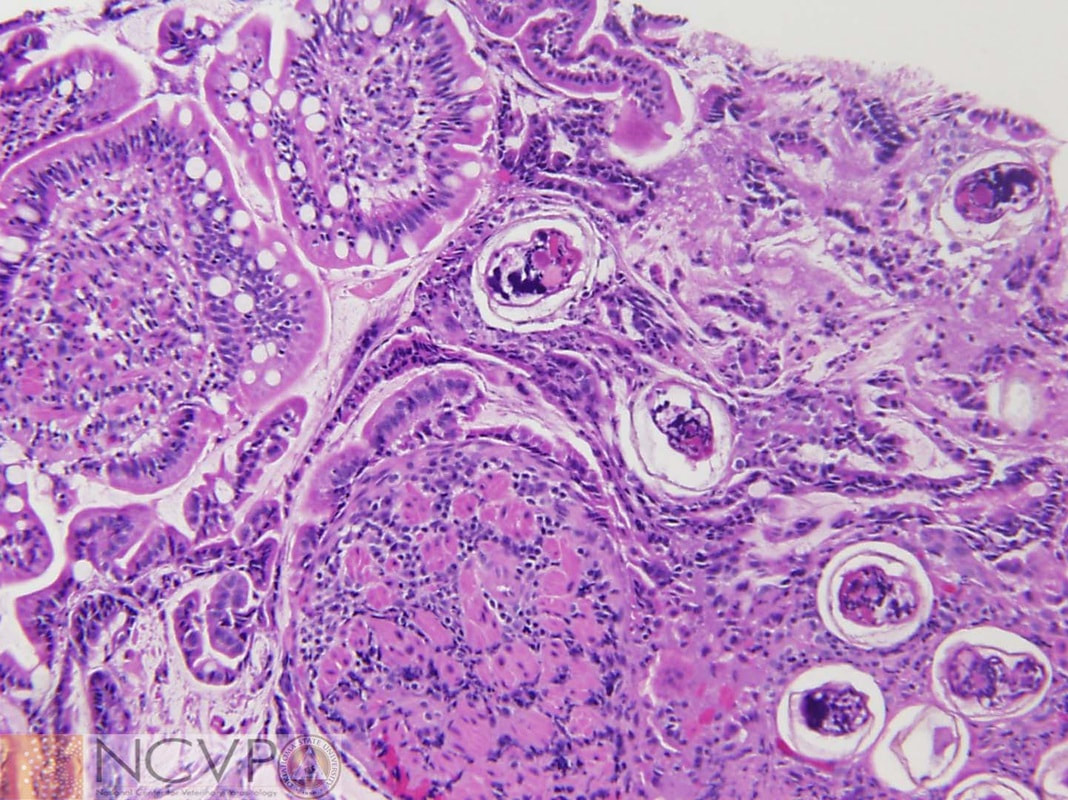
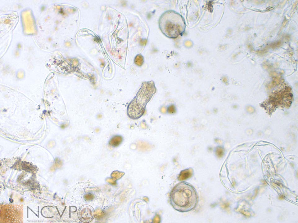
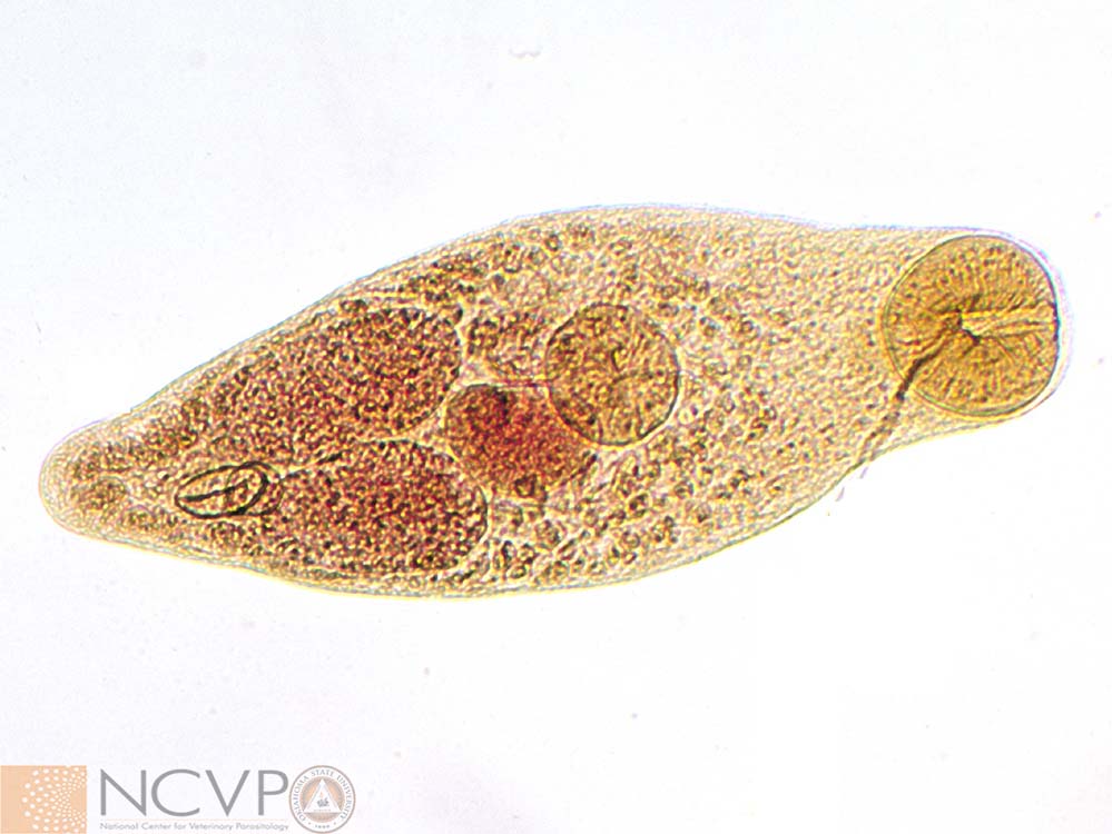
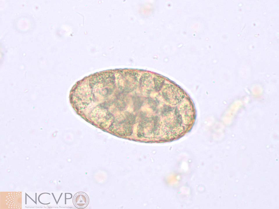
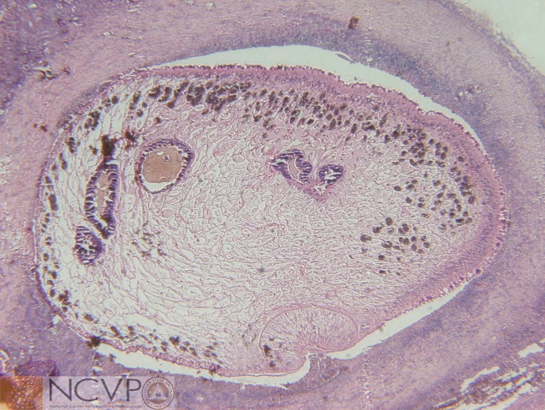
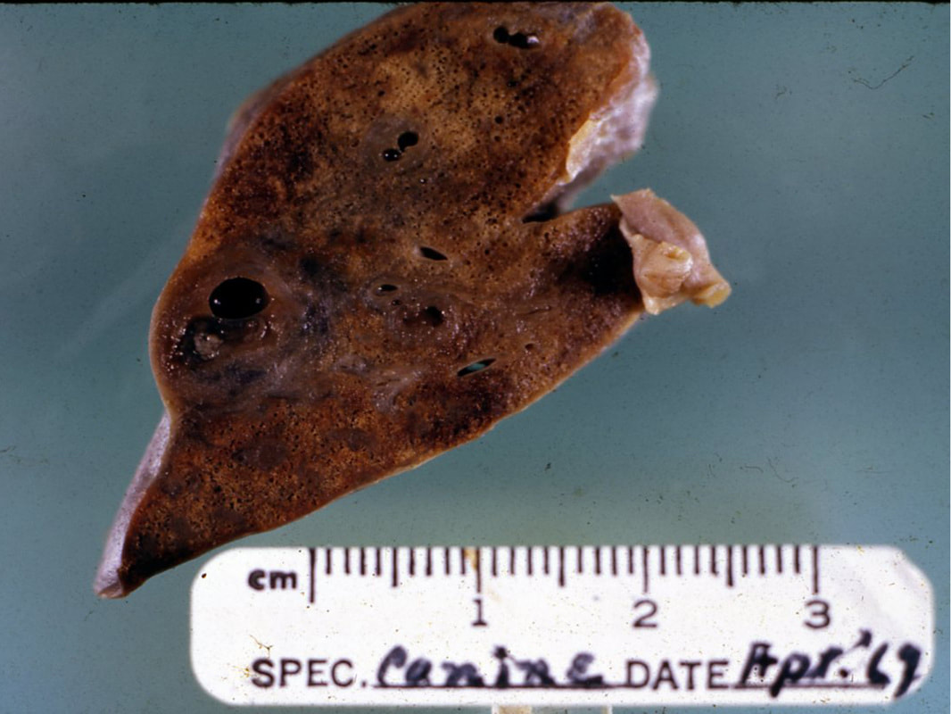
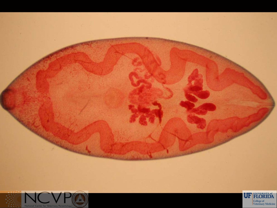
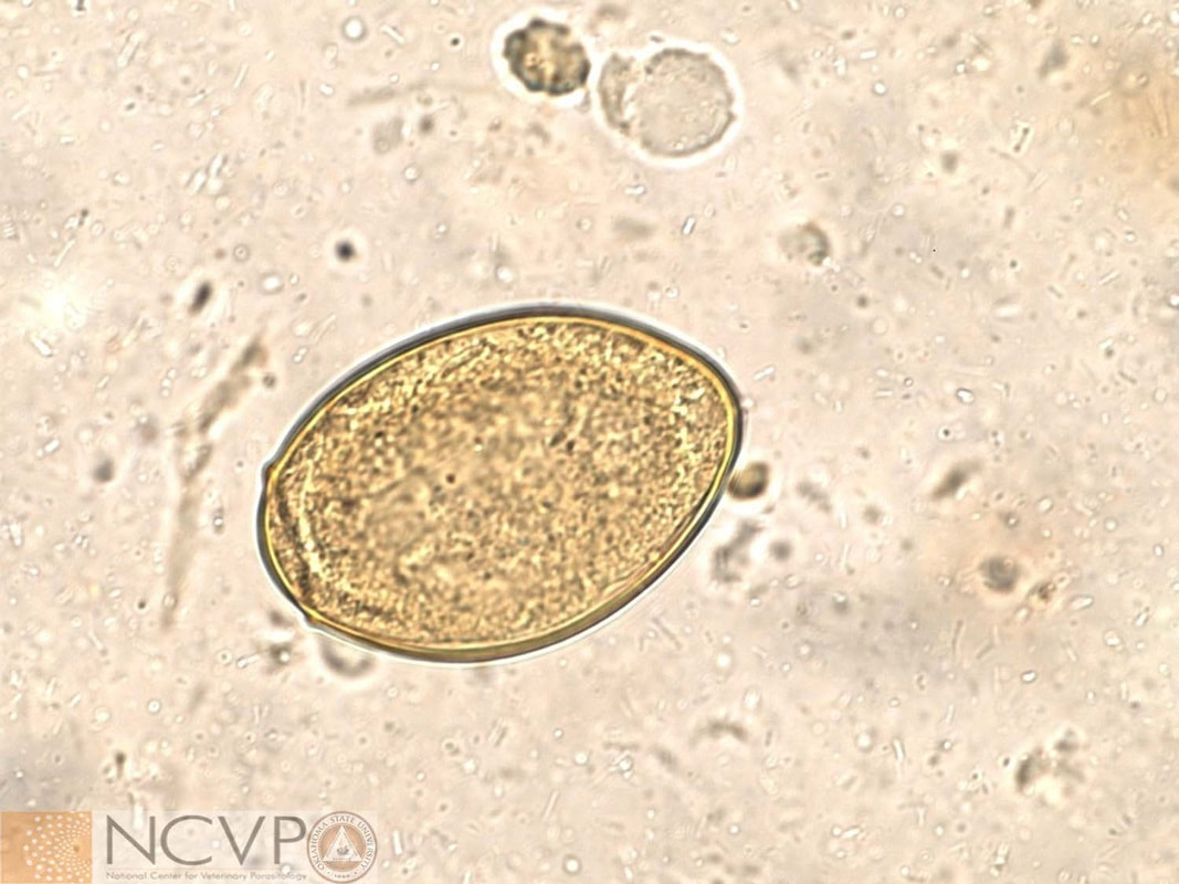
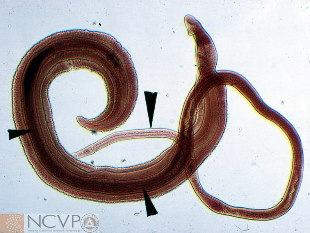
NCVP - National Center for Veterinary Parasitology

  • Home
  • Resources
    • People >
      • Directors and Advisory Board
      • Residents >
        • Boehringer Ingelheim Resident
        • Elanco Resident
        • IDEXX Resident
        • Merck Resident
        • Zoetis Resident
      • NCVP Alums
      • Clinical Parasitology Support
      • Additional Support
      • Sponsors
    • Parasite Image Database >
      • Protozoa
      • Arthropods
      • Nematodes
      • Trematodes
      • Cestodes
      • Acanthocephala
      • Tick-Borne Disease Agents
    • Case of the Month
    • Teaching and Research Materials >
      • Teaching Specimen Request
      • Parasite Jeopardy Games
      • Parasite Videos
    • Online Resources >
      • Parasitology Board Preparation
      • Additional Parasitology Resources
      • Resources for Educators
    • NCVP Board Member Resources
    • NCVP Zoom Backgrounds
    • Residents' Corner
  • News
    • Veterinary Parasitology in the News
    • NCVP Newsletter
  • Opportunities
    • Request for Proposals
    • Residency Application
    • Continuing Education in Parasitology
    • Positions Open in Veterinary Parasitology

Trematodes


Alaria spp.

Alaria sp. adult. This fluke resides in the small intestine of dogs, cats, and a variety of wild carnivores.
Eggs of Alaria sp. found in canine feces. Eggs are 98‒134 x 62‒68 µm, operculated, yellowish-brown in color, and contain an undifferentiated embryo surrounded by yolk cells.

Dicrocoelium dendriticum

Dicrocoelium dentrticum adult. This fluke infects the bile ducts of animals including domestic and wild ruminants, pigs, dogs, horses, and rabbits. This photomicrograph was taken of an original slide from the 1940s in the Dr. Wendell Krull collection.
Dicrocoelium dentriticum egg. These eggs are 38‒45 x 22‒30 µm, brownish in color, operculated, and contain a fully formed miracidium. Despite their small size, they are best recovered using fecal sedimentation procedures.

Fasciola hepatica

Adults of Fasciola hepatica recovered from the bile ducts of an infected sheep.
Adult Fasciola hepatica stained with acetocarmine to highlight internal organs.
Metacercaria of Fasciola hepatica. Metacercariae, the infective intermediate stage of Fasciola hepatica, may be found encysted on aquatic vegetation, just below the water-level. Their hard outer cyst wall allows them to survive prolonged periods in the external environment.
Cercaria of Fasciola hepatica. The cercaria has suckers and a digestive tract similar to the adult stage, along with excretory, nervous, and endocrine systems. They leave the snail intermediate host actively, and use their tails to propel themselves through water towards vegetation where they encyst.
Redia of Fasciola hepatica. This intermediate stage develops from a sporocyst within the snail. Each sporocyst produces five to eight rediae. They may then go on to produce daughter rediae under unfavorable conditions, or continue development into cercariae.
Sporocyst of Fasciola hepatica. Sporocysts are formed after miracidia penetrate soft tissues of aquatic snails. Essentially a mass of undifferentiated cells, sporocysts may be over 1 mm in length before they differentiate into rediae.
Miracidium of Fasciola hepatica. The miracidium hatch from the egg when exposed to the appropriate warm temperature, moisture, light, and salinity. Using cilia that cover its ectoderm, the miracidium will swim to find an appropriate host and bore its way into the aquatic snail, shedding its cilia and becoming a sporocyst as it enters.
Egg of Fasciola hepatica recovered using fecal sedimentation on feces from an infected cow. Eggs are typically brown in color, measure 130‒150 by 63‒90 µm, and have an operculum.
Lymnea sp. and Pseudosuccinea sp.
Lymnea on and Pseudosuccinea near a dime. These snails serve as intermediate hosts for Fasciola hepatica and their presence in a pasture is a strong indicator cattle may be at risk for infection.

Fascioloides magna

Fascioloides magna adults. Also known as the giant liver fluke or large American liver fluke, mature worms measure 4‒10 cm in length x 2‒3.5 cm in width.
Fascioloides magna adult stained with acetocarmine to highlight internal organs.
Adult Fascioloides magna in situ from a partially dissected, infected deer liver at necropsy. Fascioloides magna may infect wild ruminants, cattle, sheep, and pigs. However, wild ruminants, especially white-tailed deer, are definitive hosts and maintain F. magna cycles in wild environments. Infections of F. magna in domestic animals do not become patent and the parasite is either completely encapsulated or migrate through liver parenchyma until the infected host dies or is euthanized.

Heterobilharzia americana

H and E stained histologic section of dog pancreas with Heterobilharzia americana adult within vessel.
H and E stained histologic section of dog intestine containing Heterobilharzia americana eggs.
Stained Heterobilharzia americana adults. This fluke is unique in that adults are separate sexes. The female resides in the gynecophoric canal of the larger-bodied male.
Heterobilharzia americana egg recovered from fecal sedimentation performed with saline. These eggs are 74‒113 by 60‒80 µm and contain fully-formed miracidia.
Heterobilharzia americana miracidium. When eggs of Heterobilharzia americana are placed in water, they are stimulated to hatch, releasing miracidia that move through water to find a suitable intermediate host.

Nanophyetus salmincola

Nanophyetus salmincola adult. This fluke resides in the small intestine of piscivorous (i.e., fish-eating) mammals, including domestic dogs and cats. This trematode is the vector of Neorickettsia helminthoeca, causative agent of Salmon poisoning disease in dogs.
Nanophyetus salmincola egg recovered in a fecal sedimentation from an infected dog. These eggs are 72‒97 x 35‒55 µm, operculated, and contain an undifferentiated embryo surrounded by yolk cells.

Paragonimus kellicotti

Hemotoxlyn Eosin stained (H and E) histologic section of cat lung with adult Paragonimus kellicotti. Note the oral sucker drawing in a plug of host lung tissue.
Lung nodule in a dog induced by Paragonimus kellicotti. Infection with Paragonimus kellicotti is a result of eating an infected crayfish, or ingesting metacercariae released from an injured or degenerating crustacean.
Adult Paragonimus kellicotti stained with acetocarmine to highlight internal organs. This fluke resides in the lung parenchyma of cats, dogs, pigs, goats, and a variety of wild mammals.
Egg of Paragonimus kellicotti. Eggs are between 75‒118 x 42‒67 µm and have a thickened ridge along the line of the operculum.

Paramphistomum cervi

Paramphistomum cervi adults shown attached to the rumen wall of a cow. Paramphistomum has both an anterior and posterior sucker and the genital pore opens ventrally. Pathogenesis is due to migration of the immature fluke from the duodenum to the rumen, presenting as hemorrhagic enteritis. ​
Paramphistomum cervi adult. Termed a conical fluke, because of its pear-shaped, slightly curved shape, its large posterior subterminal sucker is easily visible. Adults may reach a size of 5‒13 mm in length and 2‒5 mm in width.
Paramphistomum cervi egg. These eggs are operculated and approximately 114‒175 by 65‒100 µm. Eggs of P. cervi overlap in size with Fasciola hepatica eggs. However, P. cervi eggs have a clear shell wall compared to those of F. hepatica which are brown or amber in color.

Platynosomum fastosum

Platynosomum fastosum adult. This liver fluke infects the bile and pancreatic ducts of cats.
Platynosomum fastosum egg from feline feces. These eggs are 34‒50 X 20‒35 µm, operculated, and best recovered by fecal sedimentation procedures.

Schistosoma

Schistosoma sp.
Adults of the genus Schistosoma. The black arrow heads indicate the location of the female Schistosoma, her posterior end is within the male's gynecophoric canal. ​

    Categories

    All